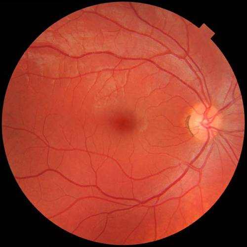
The fundus of the eye is the interior surface of the eye opposite the lens and includes the retina, optic disc, macula, fovea, and posterior pole. The fundus can be analyzed by ophthalmoscopy and/or fundus photography. The term fundus may likewise be inclusive of Bruch’s membrane and the choroid.
Types of a Fundus of the Eye
The color of the fundus differs both between and within types. In one study of primates the retina is blue, green, yellow, orange, and red; just the human fundus (from a lightly colored blonde person) is red. The major distinctions kept in mind among the “higher” primate species were size and consistency of the border of macular area, shapes and size of the optic disc, evident ‘texturing’ of retina, and coloring of retina.
Vision Support: Quality AREDS 2 Formula eye vitamins can be found on Amazon.
What Does Funduscopy Show
Medical signs that can be found from observation of eye fundus (normally by funduscopy) include hemorrhages, exudates, cotton wool spots, blood vessel problems (tortuosity, pulsation and new vessels) and coloring. Arteriolar constriction, seen as “silver wiring”, and vascular tortuosities are seen in hypertensive retinopathy.
The eye’s fundus is the only part of the human body where the microcirculation can be observed directly. The size of the capillary around the optic disc has to do with 150 μm, and an ophthalmoscope allows observation of blood vessels with diameters as little as 10 μm.