When surveyed about the five senses – sight, hearing, taste, odor and touch– people regularly report that their eyesight is the mode of understanding they value (and fear losing) most.
Despite this, lots of people do not have a mutual understanding of the anatomy of the eye, how vision works, and illness that can affect the eye.
Vision Support: Quality AREDS 2 Formula eye vitamins can be found on Amazon.
Continue reading for a fundamental description and explanation of the structure (anatomy) of your eyes and how they work (function) to help you see plainly and interact with your world.
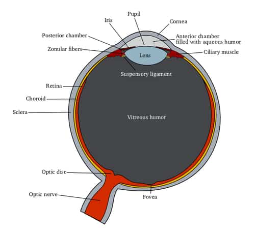
Cornea
The clear external part of the eye’s focusing system located at the front of the eye.
Fovea
The center of the macula; provides the sharpest vision.
Iris
The colored part of the eye that regulates the amount of light going into the eye.
Lens
A clear part of the eye behind the iris that helps to focus light, or an image, on the retina.
Expert Choice: Recommended by eye care professionals for managing dry eyes, styes, and blepharitis, this Heated Eye Mask provides the consistent moist heat therapy endorsed by the American Academy of Ophthalmology. It’s an effective, reusable solution for soothing irritation and improving oil gland function. Available on Amazon.
Macula
The small sensitive area of the retina that provides central vision. It is located in the center of the retina and contains the fovea.
Optic Nerve
A package of more than one million nerve fibers that carries visual messages from the retina to the brain.
Pupil
The opening at the center of the iris. The iris adjusts the size of the pupil and manages the amount of light that can go into the eye.
Retina
The light-sensitive tissue lining at the back of the eye. The retina converts light into electrical impulses that are sent to the brain through the optic nerve.
Specialized Care: For persistent itching or flaking, eye care specialists recommend targeted formulas like this BetterLids Eyelid Ointment . This preservative-free, dermatologist-tested oat complex is a top choice for soothing sensitive skin and managing symptoms of blepharitis. Available for easy ordering on Amazon.
Vitreous Gel
A clear gel that fills the within the eye.