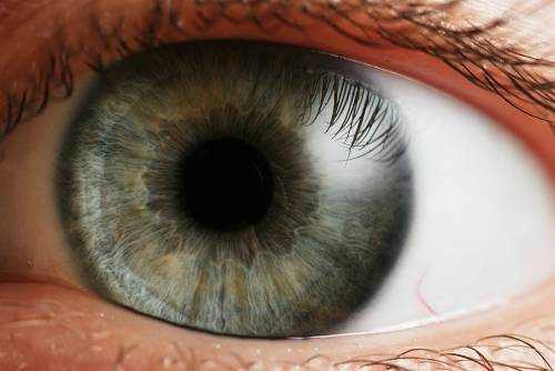
It is quite interesting to know what is the function of human iris. The iris (plural: irides or irises) is a thin, circular structure in the eye, responsible for controlling the diameter and size of the pupil and thus the amount of light reaching the retina. Eye color is specified by that of the iris. In optical terms, the pupil is the eye’s aperture, while the iris is the diaphragm.
Iris Function and Structure
The iris includes two layers: the front pigmented fibrovascular referred to as a stroma and, beneath the stroma, pigmented epithelial cells.
The stroma links to a sphincter muscle (sphincter pupillae), which contracts the pupil in a circular movement, and a set of dilator muscles (dilator pupillae) which pull the iris radially to increase the size of the pupil, pulling it in folds. The back surface area is covered by a greatly pigmented epithelial layer that is two cells thick (the iris pigment epithelium), but the front surface area has no epithelium. This anterior surface area projects as the dilator muscles. The high pigment material obstructs light from passing through the iris to the retina, restricting it to the pupil. The external edge of the iris, referred to as the root, is attached to the sclera and the anterior ciliary body. The iris and ciliary body together are called the anterior uvea. Simply in front of the root of the iris is the region referred to as the trabecular meshwork, through which the aqueous humour continuously drains out of the eye, with the outcome that diseases of the iris frequently have important results on intraocular pressure and indirectly on vision. The iris along with the anterior ciliary body supply a secondary path for liquid humour to drain from the eye.
Vision Support: Quality AREDS 2 Formula eye vitamins can be found on Amazon.
The iris is divided into two major regions:
- The pupillary zone is the inner region whose edge forms the boundary of the pupil.
- The ciliary zone is the rest of the iris that reaches its origin at the ciliary body.
The collarette is the thickest area of the iris, separating the pupillary portion from the ciliary part. The collarette is a rudiment of the coating of the embryonic pupil. It is normally specified as the region where the sphincter muscle and dilator muscle overlap. Radial ridges extend from the periphery to the pupillary zone, to supply the iris with blood vessels. The root of the iris is the thinnest and most peripheral.
The muscle cells of the iris are smooth muscle in mammals and amphibians, however are striated muscle in reptiles (consisting of birds). Numerous fish have neither, and, as an outcome, their irides are not able to dilate and agreement, so that the pupil constantly stays of a repaired size.
Anterior Surface
- The crypts of Fuchs are a series of openings located on either side of the collarette that enable the stroma and deeper iris tissues to be bathed in liquid humor. Collagen trabeculae that surround the border of the crypts can be seen in blue irises.
- The midway between the collarette and the origin of the iris. These folds result from modifications in the surface of the iris as it dilates.
- Crypts on the base of the iris are additional openings that can be observed near to the outer part of the ciliary part of the iris.
Posterior Surface
- The radial contraction folds of Schwalbe are a series of very great radial folds in the pupillary portion of the iris extending from the pupillary margin to the collarette. They are associated with the scalloped look of the pupillary ruff.
- The structural folds of Schwalbe are radial folds extending from the border of the ciliary and pupillary zones that are much more comprehensive and more commonly spaced, constant with the “valleys” between the ciliary processes.
- Some of the circular contraction folds are a fine series of ridges that run near the pupillary margin and vary in density of the iris pigment epithelium; others are in ciliary part of iris.
Iris Histology
From anterior (front) to posterior (back), the layers of the iris are:
- Anterior limiting layer
- Stroma of iris
- Iris sphincter muscle
- Iris dilator muscle (myopeithelium).
- Anterior pigment epithelium.
- Posterior pigment epithelium.
The iris of the eye structure
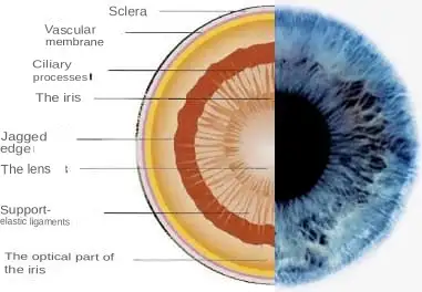
The iris has to do with 0.008 inches (0.2 mm) thick. It is thinnest at the border with the ciliary body. It is in this area that the organ might tear and bleed profusely into the eye chambers.The posterior part is adjacent to the surface area of the lens. Therefore, synechiae – blend of the lens capsule and iris pigment cells – might form during inflammatory occasions.
Expert Choice: Recommended by eye care professionals for managing dry eyes, styes, and blepharitis, this Heated Eye Mask provides the consistent moist heat therapy endorsed by the American Academy of Ophthalmology. It’s an effective, reusable solution for soothing irritation and improving oil gland function. Available on Amazon.
Iris Development
The stroma and the anterior border layer of the iris are originated from the neural crest, and behind the stroma of the iris, the sphincter pupillae and dilator pupillae muscles along with the iris epithelium develop from optic cup neuroectoderm.
The iris is normally strongly pigmented, with the color typically ranging in between brown, hazel, green, gray, or blue. Occasionally, the color of the iris is because of a lack of pigmentation, as in the pinkish-white of oculo-cutaneous albinism, or to obscuration of its pigment by blood vessels, as in the red of an unusually vascularised iris. Regardless of the large range of colors, the only pigment that contributes considerably to normal human iris color is the dark pigment melanin. The amount of melanin pigment in the iris is one factor in identifying the phenotypic eye color of an individual.
Structurally, this substantial particle is just a little various from its comparable found in skin and hair. Iris color is because of variable amounts of eumelanin (brown/black melanins) and pheomelanin (red/yellow melanins) produced by melanocytes. More of the former is discovered in brown eyed people and of the latter in blue and green-eyed people.
Genetic and Physical Factors Determining Iris Color
Iris color is a highly complicated phenomenon consisting of the combined impacts of texture, coloring, fibrous tissue and blood vessels within the iris stroma, which together comprise an individual’s epigenetic constitution in this context. An individual’s “eye color” is in fact the color of one’s iris, the cornea being transparent and the white sclera completely outside the area of interest.
Specialized Care: For persistent itching or flaking, eye care specialists recommend targeted formulas like this BetterLids Eyelid Ointment . This preservative-free, dermatologist-tested oat complex is a top choice for soothing sensitive skin and managing symptoms of blepharitis. Available for easy ordering on Amazon.
Melanin is yellowish-brown to dark brown in the stromal pigment cells, and black in the iris pigment epithelium, which lies in a thin however very opaque layer throughout the back of the iris. A lot of human irises also show a condensation of the brownish stromal melanin in the thin anterior border layer, which by its position has an overt influence on the overall color. The degree of dispersion of the melanin, which is in subcellular bundles called melanosomes, has some influence on the observed color, however melanosomes in the iris of human beings and other vertebrates are not mobile, and the degree of pigment dispersion can not be reversed. Unusual clumping of melanosomes does happen in disease and might result in irreversible changes in iris color (see heterochromia, listed below).
Colors besides brown or black are because of selective reflection and absorption from the other stromal parts. In some cases lipofuscin, a yellow “wear and tear” pigment, also enters into the visible eye color, particularly in aged or unhealthy green eyes.
How many layers are there in the iris and what are their functions?
The iris is disc-shaped and includes three layers: anterior border layer, middle stromal layer (from mesoderm), and posterior pigment-muscular layer (from ectoderm). The anterior layer is formed by connective tissue cells, under which pigment-containing cells (melanocytes) lie. Under them even deeper (in stroma) there is a network of capillaries and collagen fibers.
The posterior iris layer includes muscles – a circular sphincter of the pupil and a radially organized dilator.
The anterior iris surface area is usually divided into two belts: pupillary and ciliary. The border in between them is a circular roller, the mesentery. In the pupillary belt there is a sphincter of the student, and in the ciliary (ciliary) belt there is a dilator.The external area of the organ has lacunas or crypts, which lie between the vessels.
Abundant blood supply to the iris is provided by two posterior and several anterior ciliary arteries, which form a large arterial circle. From the latter, branches of vessels depart radially, forming a small arterial circle on the border of the pupillary and ciliary belts. The organ gets sensory innervation from long ciliary nerves that form a thick plexus.
Iris color
Iris pigmentation depends upon the number of pigment cells (melanocytes) in the stroma. Brown is a dominant characteristic, while blue is recessive.
The newborn has no melanocytes, however in the first couple of months (and years) they gradually appear, and the iris color changes. In albinos, the iris is pink in color.
In some cases, there might be a non-symmetrical distribution of pigment cells in both eyes, leading to heterochromia.
Stromal melanocytes are the source of eye melanoma development.