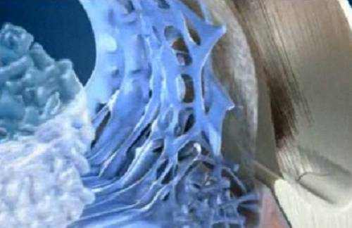
The trabecular meshwork is an area of tissue in the eye located around the base of the cornea, near the ciliary body, and is responsible for draining the liquid humor from the eye through the anterior chamber (the chamber on the front of the eye covered by the cornea).
The tissue is spongy and lined by trabeculocytes; it enables fluid to drain pipes into a set of tubes called Schlemm’s canal flowing into the blood system.
Vision Support: Quality AREDS 2 Formula eye vitamins can be found on Amazon.
Trabecular Meshwork Structure
The meshwork is divided up into three parts, with characteristically various ultrastructures:
- Inner uveal meshwork – Closest to the anterior chamber angle, includes thin cord-like trabeculae, orientated primarily in a radial style, enclosing trabeculae spaces larger than the corneoscleral meshwork.
- Corneoscleral meshwork – Contains a large amount of elastin, organized as a series of thin, flat, perforated sheets organized in a laminar pattern; considered the ciliary muscle tendon.
- Juxtacanalicular tissue (also called the cribriform meshwork) – Lies immediately surrounding to Schlemm’s canal, composed of connective tissue ground substance filled with glycoaminoglycans and glycoproteins. This thin strip of tissue is covered by a monolayer of endothelial cells.
The trabecular meshwork is helped to a little degree in the drainage of liquid humour by a second outflow pathway, the uveo-scleral pathway (5-10% of outflow happens in this manner). The uveo-scleral pathway is increased with making use of glaucoma drugs such as prostaglandins (e.g., Xalatan, Travatan).
The trabecular meshwork had previously been thought to emerge from a point (pinnacle) representing the termination of the DM (Schwalbe’s line) nevertheless it is now considered to extend into the cornea, forming the Dua’s layer.
Function of Trabecular Meshwork
Glaucoma: Glaucoma is believed to be triggered by an increase in intraocular pressure. Pressure increases either when excessive liquid humor fluid is produced or by decreased aqueous humor outflow. The trabecular meshwork is accountable for the majority of the outflow of aqueous humour.
Bottom Line
The Trabecular Meshwork has been the recent focus of surgical innovation such as ab interno trabeculectomy and the iStent. Results thus far have been restricted. More elucidating the mechanics of TM outflow such as segmental outflow will be necessary to determining the pathophysiologic basis of main open-angle glaucoma in addition to increasing the success rates of TM bypass procedures. Although investigators have begun to describe outflow physiology in the nonglaucomatous eye, the molecular pathways responsible for the pathologic modifications leading to ocular hypertension and subsequent glaucoma stay elusive.