There are two basic types of macular degeneration: “wet” and “dry.” Around 10-15% of the cases of macular degeneration are the “wet” (exudative) type.
Wet age-related macular degeneration is an eye disease that causes blurred vision or a blind spot in your visual field. It’s typically brought on by abnormal capillary that leak fluid or blood into the macula (MAK-u-luh). The macula is in the part of the retina accountable for central vision.
Vision Support: Quality AREDS 2 Formula eye vitamins can be found on Amazon.
Wet macular degeneration is one of two types of age-related macular degeneration. The other type– dry macular degeneration — is more common and less severe. The wet type constantly begins as the dry type.
Early detection and treatment of wet age-related macular degeneration might help reduce vision loss and, in some circumstances, recover vision.
Symptoms of Wet AMD
What the Patient may see: With the “wet” type, patients may see a dark spot (or spots) in the center of their vision due to blood or fluid under the macula. Straight lines may look wavy due to the fact that the macula is not smooth. Side or “peripheral” vision is rarely impacted. However, some patients do not notice any such changes, in spite of the start of neovascularization. For that reason, regular eye evaluations are still extremely important for patients at high risk.
Two Forms of Choroidal Neovascularization
There are two types of choroidal neovascularization (CNV) that have actually been recognized, “classic” and “occult.” The traditional form is well-defined and typically results in vision that is in between 20/250 and 20/400, but it might be worse than 20/800. For eyes with the occult form, the average visual acuity is somewhat much better, between 20/80 and 20/200. Occult lesions are not well-delineated and they have less leak.
Expert Choice: Recommended by eye care professionals for managing dry eyes, styes, and blepharitis, this Heated Eye Mask provides the consistent moist heat therapy endorsed by the American Academy of Ophthalmology. It’s an effective, reusable solution for soothing irritation and improving oil gland function. Available on Amazon.
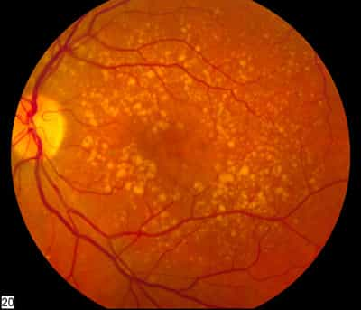
When CNV has established in one eye, whether there is a visual loss or not, the other eye is at reasonably high risk for the very same change. When all 4 risk factors — more than 5 drusen, big drusen, pigmental clumping, and systemic hypertension– are present, the five-year risk of CNV in the second eye is 87%, whereas if none of these risk factors are present, the risk is 7%.
In addition, CNV might advance quickly, and any sudden modification in main vision for that reason requires a timely assessment after dilation of the eyes. The function of this test is to find out whether the abrupt loss of vision is due to leak of blood vessels and which treatment might be suitable.