Glaucoma, often dubbed the “silent thief of sight,” is one of the leading causes of blindness worldwide. Affecting over 80 million people globally, glaucoma is a complex condition that primarily damages the optic nerve, leading to irreversible vision loss. But what exactly causes this devastating disease?
Prevalence of Glaucoma by Age Group
How Does Glaucoma Develop?
At its core, glaucoma occurs when the optic nerve, which transmits visual information from the eye to the brain, becomes damaged. The most common culprit is elevated intraocular pressure (IOP) within the eye. However, it’s not always that simple. There are forms of glaucoma where IOP remains normal, but other mechanisms still damage the optic nerve.
Vision Support: Quality AREDS 2 Formula eye vitamins can be found on Amazon.
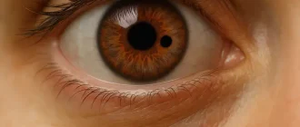
Think of the optic nerve as a high-speed internet cable. Any disruption to its integrity—whether physical or due to poor maintenance—can interfere with the signal, leading to visual impairment.
Comparison of Glaucoma Types
| Type | Prevalence (%) |
|---|---|
| Primary Open-Angle Glaucoma | 70% |
| Angle-Closure Glaucoma | 20% |
| Normal-Tension Glaucoma | 10% |
What Are the Primary Causes?
Elevated Intraocular Pressure (IOP)
Elevated IOP occurs when there’s an imbalance in the production and drainage of aqueous humor, the fluid that nourishes the eye. This pressure buildup compresses the optic nerve over time.
- Primary Open-Angle Glaucoma: The most common type, it’s often associated with slow, progressive damage due to fluid drainage issues.
- Angle-Closure Glaucoma: A rarer, more acute condition where the drainage angle is physically blocked, causing a rapid spike in pressure.
Genetic Factors
Did you know that having a family member with glaucoma increases your risk by up to 10 times? Certain genes linked to optic nerve structure and aqueous humor regulation are inherited, making genetics a significant contributor.
Role of Genetics in Glaucoma Risk
Expert Choice: Recommended by eye care professionals for managing dry eyes, styes, and blepharitis, this Heated Eye Mask provides the consistent moist heat therapy endorsed by the American Academy of Ophthalmology. It’s an effective, reusable solution for soothing irritation and improving oil gland function. Available on Amazon.
| Genetic Factor | Risk Increase (%) |
|---|---|
| Family History of Glaucoma | 80% |
| Specific Gene Mutations | 60% |
| Ethnic Predisposition | 50% |
Age and Aging
People over 60 are at a higher risk for developing glaucoma due to the natural stiffening of eye tissues and reduced fluid outflow.
Ethnic Background
African Americans, Latinos, and Asians are disproportionately affected by certain types of glaucoma. For example, open-angle glaucoma is more common in African Americans, while angle-closure glaucoma is prevalent in Asian populations.
Eye Trauma and Medical Conditions
- Blunt or penetrating injuries can disrupt the fluid drainage system or directly damage the optic nerve.
- Medical conditions like diabetes, high blood pressure, and cardiovascular disease increase the risk of optic nerve damage due to compromised blood flow.
Prolonged Use of Steroids
Long-term use of corticosteroids—whether oral, topical, or inhaled—can elevate IOP and increase glaucoma risk. If you’re using steroids for conditions like asthma or arthritis, regular eye exams are essential.
Specialized Care: For persistent itching or flaking, eye care specialists recommend targeted formulas like this BetterLids Eyelid Ointment . This preservative-free, dermatologist-tested oat complex is a top choice for soothing sensitive skin and managing symptoms of blepharitis. Available for easy ordering on Amazon.
Secondary Causes: Beyond the Usual Suspects
Some less common forms of glaucoma arise as a consequence of other eye or systemic conditions:
- Neovascular Glaucoma: Caused by abnormal blood vessel growth in the drainage angle, often due to diabetes or retinal vein occlusion.
- Pigmentary Glaucoma: Occurs when pigment granules from the iris clog the drainage system.
- Exfoliation Syndrome: Tiny flakes of material accumulate in the eye, blocking fluid outflow.
Can You Prevent Glaucoma?
While we can’t completely prevent glaucoma, early detection and intervention significantly reduce the risk of blindness. Here’s what you can do:
- Regular Eye Exams
- For individuals over 40, experts recommend comprehensive eye exams every 2-4 years.
- Those with risk factors should get checked annually.
- Manage Underlying Conditions
Keep conditions like diabetes and high blood pressure under control to minimize optic nerve stress. - Protect Your Eyes
Wear protective eyewear during high-risk activities to prevent trauma that could lead to glaucoma. - Lifestyle Choices
- Maintain a balanced diet rich in antioxidants to support eye health.
- Avoid smoking, as it negatively affects blood flow to the optic nerve.
- Engage in moderate exercise, which can lower IOP naturally.
Did You Know?
- Glaucoma often has no symptoms until advanced stages. Up to 50% of those affected are unaware they have it.
- NASA research uncovered a link between space travel and optic nerve changes. Astronauts exposed to prolonged microgravity sometimes develop increased IOP, mimicking glaucoma risk factors.
- Annual global costs of glaucoma treatment exceed $6 billion. Early diagnosis can save both vision and money.
Editorial Advice
If you’re concerned about glaucoma, prioritize regular eye exams—especially if you fall into high-risk categories. Advances in medical imaging and treatments, such as minimally invasive glaucoma surgery (MIGS), mean early intervention can effectively preserve vision. Remember, vision loss from glaucoma is irreversible, but timely action can make all the difference.
Progression of Visual Field Loss in Glaucoma