Coats disease is a telangiectasia, neovascularization disease, of unidentified aetiology, that freuquently affects unilateral eyes of kids, generally young boys. Coats disease is generally identified by unilateral (90%), progressive development of unusual vessels in the retina of the affected individuals. It’s more frequent in males (10:1) than females, and in patients younger than 8 years of ages, although it has been observed in infants along with older patients.
The disease is mainly due to aneurismatic and telangiectatic vessels as well as obstructed trichoid vessels. The typical area of these vessels is within the temporal retina. These abnormal vessels are leaking and there is exudation in numerous degrees.
Vision Support: Quality AREDS 2 Formula eye vitamins can be found on Amazon.
No genetic basis has actually been found however there has actually been explained chromosomal instability in chromosomes 3 and 13, in addition to cases in patients with Turner Syndrome (XO), Senior Loken syndrome and others.
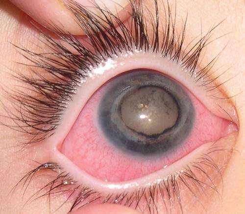
Coats disease is an rare eye condition including unusual development of blood vessels in the retina. Located in the back of the eye, the retina sends out light images to the brain and is necessary to vision. The blood-rich retinal blood vessels burst, leaking the serum portion of the blood into the back of the eye. The leak causes the retina to swell, leading to partial or total detachment of the retina. Coats disease advances gradually and impacts main vision. It is almost always unilateral (impacting only one eye). If captured early, some level of vision can typically be restored. If not dealt with until its later stages, complete loss of vision can occur. In the final stage, enucleation (elimination of the impacted eye) might be required.
What Causes Coats’ Disease?
The Etiology of the disease seems to be unknown, and there seems to be no clear predisposing factor. Nevertheless it is hypothesized that it’s due to anomalous leaking vessels. The leakage is brought on by faulty endothelium, which in turn enables blood components to flow in the retina and subretinally. This winds up in necrosis of the endothelium, abnormal neovascularization and telangiectasia.
Coats’ Disease Symptoms
Providing symptom is regularly leukocoria but the diagnosis is identified fundoscopically. In mild cases one or two focuses of retinal telangiectasia can be discovered typically on the temporal hemispheres. Microaneurysms, trichoidal obstruction and thickening of retinal venules are not uncommon, while it is possible that they are accompanied by retinal edema and exudates that may even block the pathological vessels. The exudates differ in size and they tend to occupy the inferior pole, as a result, visual acuity is reduced mostly due to seepage of the fovea, formation of cystoid macular edema or perhaps exudative retinal detachment. Those exudates ultimately cause discoid glial scarring and subretinal neo-vascularization, that can result in glaucoma and phthisis.
How Is Coats’ Disease Diagnosed?
Besides thorough fundoscopy, which eventually develops the diagnosis some clinical tests are made use of. The diagnostic tests to be used need to consist of OCT, ultrasound and fluorescence angiography.
The major scientific significance of Coats’ Disease is to differentiate it from Retinoblastoma given that both appear with leukocoria, however calcium seen in CT or ultrasound excludes Coats in favor of Retinoblastoma. Others such as Toxocara infection, retinopathy of prematurity, pars planitis, familial exudative chororetinopathy, PVF, metastatic retinitis, Norris disease, Eales disease, cavernous retinal hemangioma and leukemia, should be considered.
Expert Choice: Recommended by eye care professionals for managing dry eyes, styes, and blepharitis, this Heated Eye Mask provides the consistent moist heat therapy endorsed by the American Academy of Ophthalmology. It’s an effective, reusable solution for soothing irritation and improving oil gland function. Available on Amazon.
Differential Diagnosis
The primary medical significance of Coats Disease is to separate it from Retinoblastoma since both appear with leukocoria, but calcium seen in CT or ultrasound omits Coats in favor of Retinoblastoma. Other conditions such as Toxocara infection, retinopathy of prematurity, pars planitis, familial exudative vitreoretinopathy, retinal metastatic sores, Norrie disease, Eales disease, spacious retinal hemangioma, and leukemia, ought to be considered. A retinal vasoproliferative growth is differentiated by the presence of feeder and draining pipes vessel, an apparent vascular mass, preretinal fibrosis, and severe peripheral area.
| Feature | Coats Disease | Retinoblastoma |
|---|---|---|
| Presentation | Older patient, Unilateral, Male | Younger presentation, Maybe bilateral, Unisex |
| Leukocoria | Grey to pink | White |
| Telangiectasia | Classic, Vessels can be traced | With tumor infiltration, Vessels dip into the mass |
| Vitreous seeding | Absent | Maybe present |
| Calcification | Not a feature | Seen on USG, CT Scan |
| Intraocular mass | Not a feature | Seen in endophytic cases |
| FFA | Leaks with telangiectatic vessels | Double circulation |
Coats’ Disease Staging
A really useful staging is the one developed by Comez-Morales and that of Siegelman.
Gomez – Morales Staging
- Stage I: Focal exudates
- Stage II: Massive exudation
- Stage III: Partial exudative retinal detachment
- Stage IV: Total Retinal Detachment
- Stage V: Complications
Sigelman Staging
Specialized Care: For persistent itching or flaking, eye care specialists recommend targeted formulas like this BetterLids Eyelid Ointment . This preservative-free, dermatologist-tested oat complex is a top choice for soothing sensitive skin and managing symptoms of blepharitis. Available for easy ordering on Amazon.
- Stage I: Only telangiactasia
- Stage II: Focal exudates
- Stage III: Partial exudative retinal detachment
- Stage IV: Total Retinal Detachment
- Stage V: Complications
Coats’ Disease Treatment
The treatment of choice ranges from cryopexy or laser treatment for milder cases to a mix of both for more severe, often with the addition of scleral transplant or Vitreo-retinal surgery in basic. In addition Anti-VEGF therapy has actually been promising but safety for children is yet unidentified. Lastly enucleation of the eye is considered in painful glaucomatic eyes.
Prognosis
The prognosis depends upon the degree of the disease with milder, older presenting cases, having a better diagnosis, of even spontaneous regression, whereas children under the age of 3 have a more severe one.
People Share Their Experience
I was diagnosed with the Coats Disease when I was 4, and one of my uncles discovered that my eye would go a little to the side … my moms and dads took me instantly to see the doctor, and they identified me with the Coats Disease. Unfortunately, I had currently lost vision in my left eye.
To inform you the fact I didn’t suffer, I guess that considering that I have memory I have seen life with just one eye, my moms and dads suffered a little taking me far to see the physician, seeing how I would weep with the drops, and so on. However everything went okay, I matured; generally, absolutely nothing would tell, nor even in pictures until at 11 years of ages they started to mock me at school that it seemed that I had “mucus” stuck in my iris, thankfully I didn’t let the remarks affect me but one day playing volleyball, a dreadful unbearable pain started in my eye, I felt as if someone was squeezing it and it would come out, it injured so badly, and I likewise had a headache on the left side of my head. Instantly they took me to the physician, and they regrettably diagnosed me with glaucoma.
I went through three laser operations, and with the 3rd I was fine and currently without pain or anything like and from then up to now. I didn’t recuperate nor will I recover my sight but it is normal for me. Now I am 21, been wed for 5 and I have 4 years of ages little girl entirely healthy and without Coats disease. My pregnancy was completely normal, and without any difficulty, I only needed to go through a C-section so the blood pressure of my eye wouldn’t go up since of the birth, but I’ve had a very healthy and happy life.
If you have children or family members with this disease, let them know that they can lead a normal life like anyone’s, study, achieve their dreams, take much care of your other eye, your “good little eye” as my moms and dads would state.