A contrast sensitivity test measures your ability to distinguish between finer and finer increments of light versus dark (contrast).
This differs from common visual acuity screening in a routine eye examination, which measures your ability to acknowledge smaller sized and smaller sized letters on a basic eye chart.
Vision Support: Quality AREDS 2 Formula eye vitamins can be found on Amazon.
Contrast sensitivity is a very important procedure of visual function, especially in scenarios of low light, fog or glare, when the contrast in between objects and their background often is reduced. Owning at night is an example of an activity that needs good contrast sensitivity for safety.
Even if you have 20/20 visual skill, you can have eye or health conditions that might reduce your contrast sensitivity and make you feel that you are not seeing well.
Symptoms Of Reduced Contrast Sensitivity
If you have low contrast sensitivity, you may have problems with night owning, including trouble seeing pedestrians walking alongside badly lit streets. Or you might discover that your eyes tire more quickly while reading.
Poor contrast sensitivity likewise can increase your risk of a fall if you cannot see that you need to step down from a curb onto similarly colored pavement.
Low contrast sensitivity can be a symptom of specific eye conditions or illness such as cataracts, glaucoma or diabetic retinopathy.
Expert Choice: Recommended by eye care professionals for managing dry eyes, styes, and blepharitis, this Heated Eye Mask provides the consistent moist heat therapy endorsed by the American Academy of Ophthalmology. It’s an effective, reusable solution for soothing irritation and improving oil gland function. Available on Amazon.
Modifications in contrast sensitivity also can take place after LASIK, PRK and other types of refractive surgery.
For instance, often an individual who has LASIK might have the ability to see 20/20 after the procedure however experiences bad night vision. This could be caused by a loss of contrast sensitivity from the surgery. Conversely, some people accomplish better contrast sensitivity and night vision after LASIK, compared with their vision with glasses or contact lenses prior to the procedure.
In many cases, people with cataracts notice a significant improvement in both visual skill and contrast sensitivity after cataract surgery.
The Pelli Robson Contrast Sensitivity Testing
Contrast sensitivity screening often isn’t included in a regular eye test. Your eye doctor might perform the test since of a specific visual complaint you have or because she or he suspects you have a condition that is affecting your ability to determine contrast.
Specialized Care: For persistent itching or flaking, eye care specialists recommend targeted formulas like this BetterLids Eyelid Ointment . This preservative-free, dermatologist-tested oat complex is a top choice for soothing sensitive skin and managing symptoms of blepharitis. Available for easy ordering on Amazon.
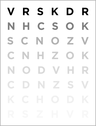
Most likely the most extensively used device to test contrast sensitivity is the Pelli Robson contrast sensitivity chart.
Other, more sophisticated devices likewise may be used to test your contrast sensitivity. These devices frequently use targets called sine-wave gratings that consist of a number of fuzzy, parallel bars of light and dark. These bars can differ in width (spatial frequency) as well as contrast from target to target, to offer a more thorough examination of how sensitive your eyes are to differences on the other hand.
Some sine-wave grating tests include a bright light source that can be directed toward your eyes during the test to mimic glare circumstances such as oncoming headlights during night-driving.
If your eye doctor identifies that you require a contrast sensitivity test, it likely will be administered after a standard visual skill test and before your pupils are dilated.
The testing generally is done while you wear your glasses or contact lenses if you require vision correction.
For assessment of eye disease, contrast sensitivity generally is evaluated on each eye separately.
For other factors, such as sports vision screening or to assess vision after contact lens fitting, LASIK or cataract surgery, the screening might be finished with both eyes open.
Contrast Sensitivity Function (CSF)
Comprehensive contrast sensitivity measurements that include both size (spatial frequency) and contrast are used to outline a person’s contrast sensitivity function (CSF).
Sine-wave grating targets with thicker bars represent low spatial frequencies; targets with thinner bars represent higher spatial frequencies. In this regard, determining an individual’s CSF is much like evaluating the sensitivity of his or her hearing, which involves using tones of low and high pitch as well as variations in volume.
Your contrast sensitivity function essentially is a plotting of the curve that defines the most affordable contrast level that you can spot for each spatial frequency evaluated.
Usually, things with high spatial frequencies (sine-wave gratings with extremely thin bars) should have considerably greater contrast than objects with lower spatial frequencies (gratings with medium-width bars) to be detected by the human visual system.
What Can Be Done About Low Contrast Sensitivity?
Your contrast sensitivity test results can assist your eye doctor figure out if you have vision errors referred to as higher-order aberrations or some other problem that might be corrected with unique glasses or eye surgery.
If you are detected with low contrast sensitivity, your optometrist might encourage you to wear corrective lenses with a yellow filter to improve your ability to discern contrast.
Likewise, glasses with customized wavefront lenses often can improve contrast sensitivity.
In some cases, custom or wavefront LASIK can minimize higher-order aberrations and improve contrast sensitivity.
Specific premium intraocular lenses (IOLs) that have actually been developed with wavefront innovation also can decrease higher-order aberrations and improve contrast sensitivity after cataract surgery.