The corneal topography test is an advanced diagnostic tool that uses specialized computer technology to create a three-dimensional map of the shape and curvature of the cornea’s surface. This test is non-invasive and painless, and it provides detailed information about various eye conditions and irregularities such as changes in corneal power, swelling, and distortions. By monitoring eye diseases and planning surgeries based on the results of this test, eye doctors are able to provide the best possible treatment for their patients without any discomfort or contact with the eye.
Importance of corneal topography test for eye care
The corneal topography examination is a necessary method of diagnosis that generates a three-dimensional depiction of the cornea’s surface curvature. This test offers a thorough understanding of the shape and strength of the cornea, which aids in identifying, monitoring, and treating various eye issues. Additionally, it is beneficial in the process of selecting appropriate contact lenses or surgical planning, specifically those for laser vision correction. Furthermore, this examination can identify abnormal conditions that are often not seen in traditional tests, making it a valuable tool for eye care professionals. The examination is also speedy, painless, and non-invasive while still providing essential details about eye health.
Purpose of Corneal Topography Test
Detecting eye conditions
The use of corneal topography can be highly beneficial in identifying eye conditions that may not be detectable through regular testing techniques. This diagnostic method provides a comprehensive visual representation of the cornea’s strength and structure, which eye specialists can use to identify and track eye diseases. It can additionally help with fitting contact lenses and preparing for surgical procedures, including laser vision correction. Moreover, corneal topography analysis can be used to assess illnesses or harm to the cornea, such as injuries or infections that may alter the cornea’s shape and impact eyesight.
Vision Support: Quality AREDS 2 Formula eye vitamins can be found on Amazon.
Mapping the cornea’s surface
A corneal topography test offers a crucial advantage of mapping the surface of the cornea. It enables eye care experts to detect and handle different cornea-related eye conditions. Similar to a topographical map depicting land surface features, these maps provide extensive information including changes in altitude and depth. By obtaining a 3D map of the cornea surface, eye care practitioners can offer precise diagnosis and customized treatment options. This leads to better visual clarity and healthier eyes for patients.
How does Corneal Topography Test Work?
Tools used in the test
The process of corneal topography involves utilizing different instruments to map out the surface of the cornea with great precision. Placido discs and scanning slit technology are the two most commonly used tools for this purpose. Placido discs work by projecting a set of circles onto the cornea, while scanning slit technology involves using a small beam of light to create an in-depth visual depiction. Through both of these methods, medical professionals can gain an accurate understanding of the cornea’s shape and curvature, which is crucial for identifying and treating various eye ailments.
Procedure for performing Corneal Topography Test
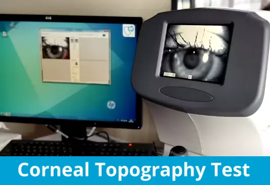
In the Corneal Topography Test, a patient faces a lit bowl containing ring patterns while data points are collected. The patient rests their head against a bar while software converts the data into a color-coded image of the cornea’s shape and power. This painless, non-invasive test provides detailed information on the corneal surface, helping doctors diagnose, monitor, and treat eye conditions such as fitting contacts and surgical planning.
Types of Corneal Topography Test
Placido discs
In case you’re curious about the purpose and function of a Placido disc in the field of eye care, you need not search any further. These discs comprise of interring circles that are illuminated on the cornea to evaluate its shape and curvature. By examining the reflection from these rings, experts in eye care can generate three-dimensional maps of the cornea and diagnose any abnormalities or diseases present. Owing to its efficacy, Placido disc technology is widely adopted in corneal topography examinations alongside scanning slit technology and Scheimpflug imaging.
Scanning slit technology
For those wanting to know more about scanning slit technology, here is a brief introduction. Scanning slit technology is a technique used to measure the curvature and strength of the cornea. It works by projecting multiple slits onto the cornea, with the camera capturing the triangulation between the reference and reflected beams to analyze its curvature and thickness. This method is useful for detecting signs of keratoconus in the early stages, and can also help track its progression.
Expert Choice: Recommended by eye care professionals for managing dry eyes, styes, and blepharitis, this Heated Eye Mask provides the consistent moist heat therapy endorsed by the American Academy of Ophthalmology. It’s an effective, reusable solution for soothing irritation and improving oil gland function. Available on Amazon.
How to read corneal topography maps
Learning how to read corneal topography maps may seem daunting at first, but it’s an essential skill for eye care professionals. These color-coded maps show the curvature and elevation of your cornea, including areas that are steep or flat. The warm colors indicate steep areas, while cool colors represent flatter areas. Another map compares your cornea to a best fit reference, tracking elevation. With these maps, eye care specialists can diagnose and manage various eye conditions, plan for surgery, and fit contact lenses to the shape of your eye.
Average cost of corneal topography test
The cost of a corneal topography test can vary depending on the location and provider. In the US, it can range from $50 to $150 per eye. While it may seem like an additional expense, the value of the information gained from this test can be invaluable in detecting and managing various eye conditions. It is important to discuss any concerns about coverage and cost with your eye care provider before having the test done.