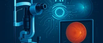
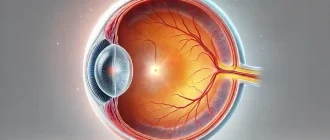
Optical coherence tomography (OCT) is revolutionizing the medical field with its ability to provide high-resolution, cross-sectional images of biological tissues. Often compared to an “optical ultrasound,” this technology uses light waves instead of sound waves to create detailed visualizations. But what exactly makes OCT so impactful, and how is it shaping the future of diagnostics?
Growth of OCT Applications Across Medical Specialties
What Is Optical Coherence Tomography?
At its core, OCT is a non-invasive imaging technique that employs low-coherence light to capture micrometer-scale images. It operates on the principle of interferometry, measuring the delay and intensity of light waves reflected from tissue structures. OCT excels in visualizing fine details, making it an indispensable tool in various medical specialties, from ophthalmology to cardiology.
How Does OCT Work?
- Emission of Light: A light source emits a low-coherence beam. This beam is finely tuned to ensure precision, allowing it to capture subtle variations in tissue structure.
- Interference Pattern Creation: The light splits into two paths—one directed at the tissue and the other at a reference mirror. When the light reflects back from these surfaces, their waves combine to form interference patterns. Think of it as watching ripples in a pond merge; these patterns contain valuable information about the tissue’s depth and texture.
- Image Reconstruction: Sophisticated algorithms process these interference patterns to generate a detailed, cross-sectional image. For example, when examining the retina, OCT provides a layered map that can distinguish between healthy and damaged tissues with micrometer precision.
This method allows clinicians to see structures beneath the surface with incredible clarity, akin to peeling back layers without physically cutting into the tissue. Imagine trying to inspect the pages of a closed book without opening it—OCT makes it possible.
Timeline of OCT Technology Advancements
| Year | Advancement |
|---|---|
| 1991 | Introduction of OCT in ophthalmology by Huang et al., providing first cross-sectional retinal images. |
| 2002 | Development of spectral-domain OCT (SD-OCT), significantly increasing image resolution and acquisition speed. |
| 2010 | Emergence of intravascular OCT for cardiology, enabling detailed coronary artery imaging. |
| 2018 | Integration of AI with OCT systems for automated retinal disease detection. |
| 2023 | Development of portable OCT devices, increasing accessibility in remote areas. |
Applications of OCT in Medicine
1. Ophthalmology: The Pioneering Field
OCT has transformed eye care, particularly in diagnosing and managing retinal diseases like macular degeneration, diabetic retinopathy, and glaucoma. For instance:
- Case Study: A 62-year-old woman from Austin, Texas, had her sight saved when OCT detected early-stage macular edema during a routine checkup, prompting timely intervention.
2. Cardiology: A Deep Dive Into Blood Vessels
Intravascular OCT helps cardiologists visualize coronary arteries to assess plaque buildup and guide stent placements. Compared to traditional angiography, OCT offers superior detail, especially for identifying smaller plaques that may not be visible otherwise. A study in Cleveland, Ohio, demonstrated how OCT-guided stent placements reduced complications by 20% compared to angiography alone.
3. Dermatology:
Emerging applications include evaluating skin lesions and early detection of melanoma. Dermatologists in Los Angeles are using OCT to monitor the progression of atypical moles without invasive biopsies, reducing patient anxiety.
4. Other Fields:
Dentistry, oncology, and even neurology are exploring OCT’s potential to uncover hidden pathologies. For example, researchers in Boston are using OCT to identify early neural changes associated with Alzheimer’s disease.
Leading Manufacturers of OCT Systems
The success of OCT technology owes much to advancements by industry leaders who design and manufacture these cutting-edge devices. Some renowned players include:
- Carl Zeiss Meditec: Known for its CIRRUS® HD-OCT systems, widely used in ophthalmology.
- Topcon Corporation: Offers innovative OCT solutions such as the Maestro2, which integrates automated scanning.
- Heidelberg Engineering: Developer of the Spectralis® OCT, which combines multimodal imaging with exceptional resolution.
- Optovue (Now Part of Visionix): Famous for the Avanti and Solix OCT systems, featuring AI-enhanced analytics.
These companies continue to push the boundaries, integrating artificial intelligence (AI) and cloud-based data analysis for enhanced diagnostic capabilities. For example, Zeiss recently introduced a feature that automatically tracks changes in a patient’s eye over time, giving specialists an edge in detecting disease progression.
Did You Know?
A study published in Ophthalmology Retina revealed that OCT can detect structural changes in the retina up to four years before symptoms of glaucoma appear, allowing for earlier treatment.
(Source: American Academy of Ophthalmology)
Advantages of Optical Coherence Tomography
Non-Invasive and Painless
Patients often describe OCT as a quick and comfortable experience—”as easy as having your picture taken.” Unlike invasive diagnostic procedures that may require needles, incisions, or contrast agents, OCT relies entirely on light, ensuring a completely non-intrusive process. This makes it particularly appealing for children and elderly patients who may be sensitive to invasive methods.
High Resolution
OCT delivers images with micrometer resolution, far surpassing the capabilities of other imaging modalities like MRI or CT for specific tissues. While MRI excels in providing a comprehensive view of deeper structures and soft tissues, and CT is highly effective for imaging bones and detecting cancers, neither can match OCT’s precision for surface and near-surface layers. This makes OCT the gold standard for applications like retinal imaging, where fine details are critical. For example, a clinician in Chicago used OCT to detect a subtle tear in the retina that was missed by an MRI, potentially saving the patient from vision loss.
Diagnostic Accuracy Rates
| Method | Accuracy (%) |
|---|---|
| OCT (Optical Coherence Tomography) | 95% |
| MRI (Magnetic Resonance Imaging) | 85% |
| CT (Computed Tomography) | 75% |
| Ultrasound | 65% |
| X-Ray | 55% |
Rapid Results
Most OCT scans take only a few minutes, providing real-time insights during clinical visits. Compared to MRI scans, which can last 30 minutes to an hour and require additional time for analysis, or CT scans that involve radiation exposure and post-processing, OCT stands out for its efficiency and immediacy. Clinicians can often interpret results on the spot, enabling faster decision-making and treatment planning.
Versatility Across Disciplines
Unlike CT, which is limited by its reliance on ionizing radiation, or ultrasound, which struggles with certain tissue densities, OCT can be adapted for a wide range of uses. For instance, researchers in New York City are exploring its potential in assessing fetal development during high-risk pregnancies, an area where traditional methods face significant limitations.
Limitations of OCT
- Cost: OCT devices can be expensive, often ranging from tens of thousands to several hundred thousand dollars. This high price tag can make it difficult for smaller clinics or private practices to afford the equipment, potentially limiting access for patients in less densely populated or rural areas.
- Limited Penetration Depth: While excellent for superficial tissues, OCT struggles with imaging deeper structures, such as those located beneath denser or more opaque layers of tissue. This limitation is particularly noticeable in applications beyond ophthalmology and dermatology, where deeper imaging is essential for comprehensive diagnostics.
- Operator Skill: Effective use requires specialized training. Mastering OCT technology involves understanding its principles, operating the equipment accurately, and interpreting the results correctly. In some cases, misinterpretation by undertrained staff could lead to diagnostic errors, emphasizing the importance of thorough education and certification for operators.
What Does the Future Hold for OCT?
The integration of AI and machine learning into OCT systems promises automated diagnostics with unparalleled accuracy. For instance, AI-powered OCT devices can already analyze retinal scans to detect early signs of diabetic retinopathy without human oversight. Additionally, portable OCT devices are becoming more common, broadening accessibility. Imagine a handheld OCT scanner available in remote areas, bringing advanced diagnostics to underserved populations. With advancements like these, the future of OCT looks incredibly bright.
Editorial Advice
Optical coherence tomography has proven to be a game-changer across multiple medical disciplines. While its cost and technical complexity may pose challenges, the benefits far outweigh these limitations. If you’re considering an OCT scan, consult a specialist equipped with state-of-the-art technology—it could be a life-changing decision. As research and innovation continue, OCT’s capabilities are poised to expand even further, offering hope and precision like never before.
Patient Experience Ratings


