Ocular Cicatricial Pemphigoid is shortened OCP. Ocular Cicatricial Pemphigoid is thought about a subtype of Mucous Membrane Pemphigoid (abbreviated MMP), and these terms are in some cases used interchangeably.
What Is Ocular Cicatricial Pemphigoid
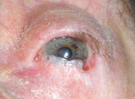
Ocular Cicatricial Pemphigoid is a kind of autoimmune conjunctivitis that leads to cicatrization (i.e. scarring) of the conjunctiva. If Ocular Cicatricial Pemphigoid is left unattended, it can cause blindness.
Vision Support: Quality AREDS 2 Formula eye vitamins can be found on Amazon.
The exact pathogenesis of Ocular Cicatricial Pemphigoid stays to be found out however the existing proof supports a Type II hypersensitivity action brought on by an autoantibody to a cell surface area antigen in the basement membrane of the conjunctival epithelium and other comparable squamous epithelia.
Examinations into the underlying target antigen have led to several possible suspects. The autoantigens accountable for bullous pemphigoid (BP230 (i.e. Bullous pemphigoid antigen I, a desmoplakin) and BP180 (i.e. Bullous pemphigoid antigen II, a transmembrane hemidesmosome)) were studied, and the sera of patients with Ocular Cicatricial Pemphigoid was shown to bind these antigens. However, more examination supports that the more likely autoantigen is in fact the beta-4 subunit of the alpha-6 beta-4 integrin of hemidesmosomes.
Research Studies of HLA (human leukocyte antigen) typing have actually discovered an increased vulnerability to the disease in patients with HLA-DR4.
What Causes Ocular Cicatricial Pemphigoid
Although the precise mechanism stays to be elucidated, the existing evidence supports the production of an autoantibody in prone people to the beta-4 subunit of the alpha-6 beta-4 integrin of hemidesmosomes in the lamina lucida of the conjunctival basement membrane.
Binding of the autoantibody to the autoantigen triggers enhance, leading to cytotoxic damage of the conjunctival membrane. Disruption of the conjunctival basement membrane subsequently results in bullae development.
Expert Choice: Recommended by eye care professionals for managing dry eyes, styes, and blepharitis, this Heated Eye Mask provides the consistent moist heat therapy endorsed by the American Academy of Ophthalmology. It’s an effective, reusable solution for soothing irritation and improving oil gland function. Available on Amazon.
The involved cellular inflammatory infiltrate of the epithelium and substantia propria manifests as the chronic conjunctivitis that is the trademark of this disease. Eosinophils and neutrophils moderate inflammation in the early and severe phases of the disease, similar to what is observed in the skin. Chronic disease has largely lymphocytic seepage.
Fibroblast activation results in subepithelial fibrosis, which in early disease appears as great white striae most easily seen in the inferior fornix. With time contract, the fibrotic striae agreement, causing conjunctival shrinkage, symblepharon formation, and forniceal shortening. In severe cases of conjunctival fibrosis, entropion, trichiasis and symblepharon may establish, leading to associated keratopathy and corneal vascularization, scarring, ulceration, and epidermalization.
The medical course and severity varies. Recurrent inflammation causes loss of Goblet cells and obstruction of lacrimal gland ductules, causing aqueous and mucous tear shortage. The resulting xerosis is severe, and together with progressive subepithelial fibrosis and destruction of limbal stem cells results in ocular keratinization.
In addition, screening of the tears of patients with Ocular Cicatricial Pemphigoid found elevated levels of IL-8, Matrix Metalloproteinase (MMP) 8, MMP-9, and myeloperoxidase (MPO), which are believed to arise from neutrophilic infiltrate in patients with Ocular Cicatricial Pemphigoid.
Specialized Care: For persistent itching or flaking, eye care specialists recommend targeted formulas like this BetterLids Eyelid Ointment . This preservative-free, dermatologist-tested oat complex is a top choice for soothing sensitive skin and managing symptoms of blepharitis. Available for easy ordering on Amazon.
Stages of Ocular Cicatricial Pemphigoid
There are a number of scientific scoring systems for Ocular Cicatricial Pemphigoid, consisting of schema from Foster, Mondino, and Tauber. Clinicians vary in which system they use for grading disease medically and although there are proponents for each system, no consensus exists regarding which system is best to use. The existing category schema are limited by the absence of direct connection with disease development and for that reason no system can be used to anticipate need for immunosuppression.
Mondino’s Classification System is based on inferior forniceal depth. A normal inferior forniceal depth is roughly 11 mm.
- Stage I: up to 25% inferior forniceal depth loss
- Stage II: 25-50% inferior forniceal depth loss
- Stage III: 50-75% inferior forniceal depth loss
- Stage IV: higher than 75% inferior forniceal depth loss
Foster’s Classification System has four stages also and is based on specific medical signs:
Stage I: Early stage
May include nonspecific symptoms and very little findings which result in under-recognition of the disease.3, 17 Commonly presents as chronic conjunctivitis, tear dysfunction, and subepithelial fibrosis. Subepithelial fibrosis manifests as great gray-white striae in the inferior fornix. Signs and symptoms are generally bilateral, and may be asymmetric.
Stage II: Shortening of the fornices
A normal inferior forniceal depth is roughly 11 mm. A reduced inferior forniceal depth is irregular and need to prompt additional examination.
Stage III: Symblepharon development
Can be spotted by pulling the lower eyelid down while the patient searches for and vice versa.
Stage IV: Ankyloblepharon
Represents end-stage disease, with surface keratinization, and substantial adhesions between the eyelid and the world, resulting in restricted motility.
Ocular Cicatricial Pemphigoid Diagnosis
Medical diagnosis is based on clinical signs and favorable direct immunofluorescence testing of the conjunctiva. Conjunctival biopsy of an actively included area is required and the conjunctival tissue needs to be sent unfixed for analysis. If participation is diffuse, biopsy of the inferior conjunctival fornix is advised. Sensible biopsy is a good idea as Ocular Cicatricial Pemphigoid is an obliterating disease of the conjunctiva and just the minimal amount of tissue necessary should be removed. Additionally, biopsy of an active oral mucosa lesion can be diagnostic too.
Immunofluorescence reveals linear staining of the epithelial basement membrane zone. The sensitivity of immunofluorescence may be as low as 50%, specifically for longstanding/severe cicatrization because of the loss of immunoreactants and the destruction of basement membrane in longstanding disease.
Serological screening is not consistently used in diagnosis. Sequential pictures work to keep an eye on medical development.
Treatment for Ocular Cicatricial Pemphigoid
Without treatment, the disease progresses in approximately 75% of patients. While systemic treatment stops progression of cicatrization in most patients, it stops working in around 10% of them. Systemic therapy is required in Ocular Cicatricial Pemphigoid as ocular participation comprises a high risk subset of MMP and is insufficiently treated with topical therapy alone. Systemic treatment is best handled by a doctor trained in the management of anti-inflammatory and immunomodulatory treatment provided the considerable risk of systemic complications requiring regular blood test tracking. A number of drugs work in dealing with Ocular Cicatricial Pemphigoid and a step-wise method of escalation of therapy when there is inadequate action is recommended.
Topical therapy can be used as an accessory for surface disease but must not be used in place of systemic therapy. Topical therapy consists of optimizing lubrication of the ocular surface with artificial tears and prompt plugging. Topical and subconjunctival steroids can alleviate symptoms however are inefficient for treatment of the underlying disease. Topical cyclosporine has been discovered to be ineffective while topical tacrolimus has actually been revealed to be effective in little case series. Subconjunctival mitomycin-c has also been examined in little case series with variable result.
If the disease remains quiescent following a few years of systemic therapy, many practitioners are frequently able to discontinue systemic therapy effectively. Nevertheless, it is essential to continue to keep track of the patient for recurrence of disease as up to 22% of patients regression.
1. Mild Disease
Dapsone is an efficient and typically used anti-inflammatory treatment in Ocular Cicatricial Pemphigoid for moderate disease and in the absence of rapid progression. Dapsone is started at a dose of 50 mg/day and gradually increased as endured by approximately 25mg every 7 days to a reliable dosage, which is generally between 100-200mg/ day. If considerable enhancement is not accomplished within 3 months, escalation of therapy is advised such as to azathioprine or methotrexate.
Systemic complications of dapsone consist of hemolysis and methemoglobinemia. G6PD (glucose-6-phosphate dehydrogenase) shortage is a contraindication to dapsone therapy as dapsone can precipitate a hemolytic crisis. All patients need to be evaluated for G6PD deficiency before initiation of therapy with dapsone.
Sulphapyridine is also an oral antibiotic and is a well-tolerated alternative in patients with mild disease who are not able to take dapsone. Sulphapyridine’s effectiveness (effective in roughly 50% of patients) is lower than dapsone nevertheless.
2. Moderate to Severe Disease
Corticosteroids have a rapid impact and work during the acute stage of severe or quickly progressive disease. Adjuvant corticosteroid-sparing immunomodulatory/immunosuppressive therapy needs to be initiated simultaneously as it might take weeks to become restorative. This will allow a quicker taper from steroids and the shortest course of steroid therapy essential given the substantial systemic side effects of long-lasting steroid therapy. Normally, one quiescence is attained, steroids are tapered slowly. Evaluating for tuberculosis (TB) is advised prior to the initiation of steroid therapy.
Azathioprine has been revealed to be an efficient steroid sparing therapy. It takes 8-12 weeks of treatment to achieve optimum impact and hence ought to be used at first concurrently with steroids. Screening for thiopurine methyltransferase (TPMT) shortage is advised prior to initiation of azathioprine as TPMT-deficient patients are at greater risk of establishing mvelosuppression. Systemic complications include leukopenia, pancytopenia, infection, malignancy, and drug-induced hypersensitivity syndrome.
Methotrexate has actually been shown to be an efficient monotherapy for Ocular Cicatricial Pemphigoid with fewer unfavorable results when compared to azathioprine, cyclophosphamide, and dapsone. The Systemic Immunosuppressive Therapy for Eye Diseases (SITE) trial discovered that cyclophosphamide was effective in managing inflammation in 70.7% of patients with Ocular Cicatricial Pemphigoid at 1 year, with 66.9% patients on less than or equal to 10mg of prednisone. Low dose methotrexate is especially reliable in mild to moderate Ocular Cicatricial Pemphigoid. Systemic complications include hepatotoxicity, nephrotoxicity, pneumonitis, lung fibrosis, pancytopenia, and malignancy.
Tetracyclines are a well-tolerated anti-inflammatory representative and have been found to be effective for mild to moderate Ocular Cicatricial Pemphigoid, especially when integrated with nicotinamide.
Mycophenolate mofetil has been revealed to be a well tolerated and reliable therapy for Ocular Cicatricial Pemphigoid. Restorative dose is usually 1000-2000mg/day. Systemic complications include leukopenia.
Cyclosporine has just been used in little series of patients and has actually been reported to have variable levels of effectiveness.
3. Severe Disease
Cyclophosphamide is first line in patients with severe disease or fast progression. It needs to be started in conjunction with steroids and can be dosed orally or IV. A short course of pulsed IV therapy (ex. 3 days) can be particularly effective in achieving rapid control if needed, such as prior to surgery. The SITE trial discovered that cyclophosphamide worked in managing inflammation in 80.8% of patients with Ocular Cicatricial Pemphigoid at 1 year, with 58.5% of patients on less than or equivalent to 10mg of prednisone. Systemic complications consist of myelosuppression, carcinogenesis, and teratogenicity.
Intravenous Immunoglobulin (IVIG) is scheduled for patients with progressive disease that is unresponsive to systemic steroids and cyclophosphamide and has been discovered to be an efficient therapy. Dosing is every 3-4 weeks till quiescence is attained, generally needing 4-12 cycles. Systemic complications are severe, and include anaphylaxis, disseminated intravascular coagulation (DIC), and intense renal failure. For that reason, IVIG is which is booked for refractory disease.
Biologics, including the anti-TNF agents Etanercept and infliximab, the IL-2 antagonist daclizumab, and the anti-CD20 antibody rituximab have actually been shown to be effective in small studies of patients with refractory Ocular Cicatricial Pemphigoid. The mix of IVIG and rituximab has actually been revealed to be effective too in refractory Ocular Cicatricial Pemphigoid.
Ocular Cicatricial Pemphigoid Complications
Seemingly minor surgical intervention and conjunctival trauma can lead to major worsening of disease. Surgical intervention, such treatment of trichiasis, entropion and cataract ought to be delayed if possible until control of active disease is achieved. In some circumstances this may not be possible and a multi-disciplinary approach is best.
Inferior eyelid retractor plication for trichiasis prevents surgery on the conjunctiva and has been revealed to be safe and efficient when undertaken in the setting scientifically quiescent Ocular Cicatricial Pemphigoid. Cryotherapy for the treatment of trichiasis has actually likewise been shown to be safe and moderately effective when undertaken in the setting medically quiescent Ocular Cicatricial Pemphigoid. In a case series of patients with well controlled Ocular Cicatricial Pemphigoid going through entropion repair, successful repair work was carried out in all patients no matter kind of surgery.
Safe and effective performance of cataract surgery has actually been shown in numerous case series of patients with well-controlled Ocular Cicatricial Pemphigoid. A clear corneal cut is suggested to reduce the risk of exacerbation.
Glaucoma is likewise a possible issue of Ocular Cicatricial Pemphigoid and is particularly tough to diagnose and treat. IOP measurements are unreliable, and examination and ancillary screening are limited by ocular surface area disease. A case series of 61 patients with severe Ocular Cicatricial Pemphigoid found that 21% of patients likewise had glaucoma and an additional 9% developed glaucoma during the follow-up.
Ocular Cicatricial Pemphigoid has actually been described in patients with other concurrent rheumatologic diseases consisting of rheumatoid arthritis, lupus, and HLA-B27 spondyloarthropathies.