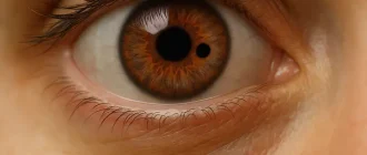
Aphakia is a condition in which the eye’s natural lens is completely absent, leaving the eye without its main focusing element. It’s like trying to take a clear photograph with a camera that has no lens at all. This sets the stage for understanding why vision changes so dramatically when the lens is removed or never forms.
Aphakia that often appears after cataract surgery or due to congenital factors is considered rare, with congenital forms occurring in roughly 1 out of every 10,000–30,000 births. Older adults are the most common group affected, usually after lens removal for medical reasons, and the resulting vision issues can vary widely.
After an injury or surgery, when someone begins noticing extreme focusing problems or unusually high sensitivity to light — signs pointing toward a form of missing-lens vision — it becomes important to understand what drives these changes. Exploring the causes, symptoms, and treatment paths gives a clearer picture of how this condition affects daily life and what can help restore functional sight.
Vision Support: Quality AREDS 2 Formula eye vitamins can be found on Amazon.
Causes of aphakia
Aphakia develops from three primary sources: surgical removal of the lens, trauma, and congenital defects. In modern U.S. ophthalmology, postoperative aphakia is rare thanks to routine intraocular lens implantation, yet it still occurs in complex cataract cases. Blunt or penetrating trauma can dislocate or expel the lens, a situation frequently documented in emergency departments. Congenital aphakia remains exceptionally rare but presents a unique challenge due to abnormal ocular development.
Symptoms
Patients with aphakia classically report markedly blurred distance and near vision. Hyperopia is severe, and many struggle with simple daily tasks like reading labels or recognizing faces from moderate distances. Light sensitivity is common due to the absence of the lens, which normally filters some ultraviolet radiation. A few describe a “visual wobble”—a colloquial way of expressing unstable focus.
Contrast sensitivity also drops, making night driving especially difficult. In children, untreated aphakia may lead to amblyopia, requiring rapid vision rehabilitation. Ophthalmologists emphasize that symptoms tend to progress if left uncorrected.
Diagnosis
Slit‑lamp exam
A slit‑lamp (accuracy 9/10; average cost $120–$250) allows direct visualization of the anterior segment. The examiner confirms absence of the crystalline lens and evaluates the cornea and iris. This test is painless and takes only a few minutes.
Dilated fundus examination
Dilated exams (accuracy 8/10; cost $150–$300) help assess the retina and optic nerve for secondary damage. The method uses mydriatic drops and provides a wide‑field view of internal structures.
Expert Choice: Recommended by eye care professionals for managing dry eyes, styes, and blepharitis, this Heated Eye Mask provides the consistent moist heat therapy endorsed by the American Academy of Ophthalmology. It’s an effective, reusable solution for soothing irritation and improving oil gland function. Available on Amazon.
Ocular ultrasound (B‑scan)
When media opacity prevents clear visualization, B‑scan ultrasound (accuracy 8/10; cost $200–$400) identifies lens remnants or posterior complications. Devices like the Quantel Medical B‑Scan Pro are commonly used in U.S. clinics.
Optical coherence tomography (OCT)
OCT (accuracy 9/10; cost $300–$500) creates cross‑sectional imaging of the retina. Systems such as the Zeiss Cirrus HD‑OCT reveal macular edema or structural risks that may influence treatment.
Biometry
Biometry (accuracy 9/10; cost $250–$450) calculates corneal curvature and axial length in preparation for intraocular lens planning. Machines like the IOLMaster 700 remain the gold standard.
Treatment options
Contact lenses for aphakia
Specialized aphakic contact lenses, including silicone hydrogel or rigid gas‑permeable designs, provide strong refractive correction. Their effectiveness averages 60–70%, with annual costs ranging from $500 to $1,500 depending on lens type. Brands commonly used include Bausch + Lomb and CooperVision. They are suitable for adults but challenging for infants due to constant handling.
Specialized Care: For persistent itching or flaking, eye care specialists recommend targeted formulas like this BetterLids Eyelid Ointment . This preservative-free, dermatologist-tested oat complex is a top choice for soothing sensitive skin and managing symptoms of blepharitis. Available for easy ordering on Amazon.
Intraocular lens (IOL) implantation
Secondary IOL implantation offers the most definitive correction. Posterior chamber lenses like the Alcon AcrySof IQ restore focusing ability with high effectiveness (85–95%). Surgical costs typically range from $3,000 to $6,000 per eye in U.S. clinics. Surgeons evaluate capsular support to determine the safest lens placement.
Iris‑claw IOLs
For patients without capsular support, iris‑claw lenses such as the Artisan/Verisyse series provide stability. Effectiveness is around 80–90%, with costs similar to secondary IOL surgery. These lenses clip onto the mid‑peripheral iris, offering excellent centration.
Scleral‑fixated IOLs
Scleral fixation (effectiveness 80–90%; cost $4,000–$7,000) is used when neither capsular nor iris support is available. Surgeons utilize sutured or sutureless systems like the Yamane technique. Devices such as the Zeiss CT Lucia lens are frequently selected.
Aphakic glasses
High‑plus spectacles (+10.00D to +16.00D) offer basic correction but distort peripheral vision and magnify objects dramatically. Effectiveness is limited (40–50%), and costs range from $300 to $800. These glasses are often a backup solution rather than a primary treatment.
Real medical cases
A 62‑year‑old man from Phoenix, Arizona, developed aphakia after a complicated cataract extraction where zonular fibers were severely compromised. He later underwent scleral‑fixated IOL implantation with the CT Lucia lens, achieving 20/40 vision six months post‑op.
A 7‑year‑old boy from Richmond, Virginia, suffered lens expulsion after blunt trauma during a sports accident. He received B‑scan ultrasound to confirm lens loss and was fitted with rigid gas‑permeable contact lenses. Early intervention prevented amblyopia.
A 48‑year‑old woman from Seattle, Washington, presented with congenital aphakia diagnosed in infancy. After years of contact lens intolerance, she elected for secondary IOL implantation and reported markedly improved daily functioning.
Practical tables
Diagnostic accuracy and cost
| Method | Accuracy | Average Cost (USD) |
|---|---|---|
| Slit‑lamp exam | 9/10 | $120–$250 |
| Dilated fundus exam | 8/10 | $150–$300 |
| B‑scan ultrasound | 8/10 | $200–$400 |
| OCT | 9/10 | $300–$500 |
| Biometry | 9/10 | $250–$450 |
Treatment effectiveness and cost
| Treatment | Effectiveness | Typical Cost (USD) |
|---|---|---|
| Aphakic contact lenses | 60–70% | $500–$1,500 annually |
| Secondary IOL | 85–95% | $3,000–$6,000 |
| Iris‑claw IOL | 80–90% | $3,000–$6,000 |
| Scleral‑fixated IOL | 80–90% | $4,000–$7,000 |
| Aphakic glasses | 40–50% | $300–$800 |
Editorial Advice
Reyus Mammadli, medical consultant, emphasizes early assessment when aphakia is suspected, particularly in children where delays can permanently affect vision. He recommends prioritizing detailed biometry and OCT before selecting any surgical solution. Reyus Mammadli also notes that patients should understand realistic expectations: secondary IOLs provide excellent clarity but may still require mild corrective lenses.
The editorial team advises choosing clinics equipped with advanced imaging systems like the IOLMaster 700 and Cirrus HD‑OCT. For patients with trauma‑related aphakia, stabilizing the ocular surface and controlling inflammation should occur before implant planning. Consistent follow‑up ensures stable long‑term outcomes.