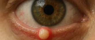
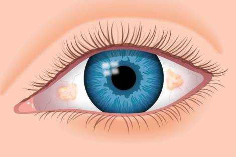
A pinguecula is a small yellow mass localized in the nasal half of the conjunctiva. Depending on its diameter, it can be latent or cause discomfort, a dry and foreign body sensation, and eye hyperemia. Diagnosis of pinguecula includes external examination, fluorescence angiography, biomicroscopy, ferning test, and OCT.
Conservative treatment is limited to the application of moisturizing drops, ointments and artificial tears. Anti-inflammatory and antibacterial therapy is indicated in the development of secondary complications. In the absence of the effect of medical treatment, surgical removal of a pinguecula is recommended.
General information
Pingvecula is a disease in ophthalmology, the main manifestation of which is the formation of a yellowish thickening on the orbital conjunctiva. The first information on this pathology dates back to 1550 BC. In the medical records of the ancient Egyptians, pinguecula was described as “fatty deposits in the eye”.
The disease can occur in isolation or as one of the manifestations of genetically determined Gaucher disease. In 95% of cases, pinguecula is diagnosed between the ages of 50 and 60. If the conjunctiva comes into prolonged contact with the factors provoking the development of this pathology, the neoplasm may develop at the age of 20-30.
Pingvecula is equally diagnosed in male and female patients. People living in a hot and dry climate are more exposed to the risk of developing the disease.
Causes of pinguecula
The etiology of pinguecula is not fully understood. As a rule, the disease develops against the background of age-related or dystrophic changes of conjunctiva. The intensity of metabolic processes decreases with age, resulting in the accumulation of products of fat and protein metabolism in the body. Reduced metabolic levels are associated with the development of pinguecula.
Also in the pathogenesis of this pathology there is degeneration of collagen fibers in the stromal part of the conjunctiva with subsequent reduction of epithelial thickness. Prolonged exposure to ultraviolet rays is a trigger for the disease. UV radiation stimulates the synthesis by fibroblasts of an abnormal fibrillar protein (elastin), which aggravates the dystrophic changes in the conjunctiva. Therefore, the risk group includes people whose work involves prolonged exposure to the sun.
Furthermore, occasional irritation of the conjunctiva by strong winds, smoke, exhaust fumes, toxic fumes, and industrial chemicals may also provoke pinguecula development.
Some experts believe that the risk of developing the pathology increases with prolonged contact lens wear. Pinguecula may form against a background of traumatic damage to the conjunctiva, scarring changes or chronic conjunctivitis.
The negative effect of infrared radiation on the development of the disease has been proved. The sclera, which is reflective, potentiates the negative effect of infrared and ultraviolet radiation on the conjunctiva and aggravates the progression of pinguecula.
Symptoms
Pinguecula in 50% of cases is a bilateral disease localized in the medial conjunctiva. There is a tendency to slow progression, which is manifested by the increase in size of the neoplasm.
The pathology is characterized by a benign course. There are no observed cases of malignant transformation of pinguecula. As a rule, patients find a small yellow area of thickening on the background of unchanged bulbar conjunctiva.
The development of the disease does not result in decrease of visual acuity, at small size it does not influence the quality of life of patients.
At the initial stages, pinguecula is characterized by a latent course. Clinical manifestations are seen when the diameter of the neoplasm increases. Patients complain of discomfort accompanied by dryness in the affected eye.
Periodic irritation of the pinguecula results in conjunctival hyperemia. Patients experience a sensation of a foreign body which provokes the development of lacrimation. In rare cases, corneal opacity is a symptom of pinguecula.
Prolonged irritation of the thickened area of the conjunctiva will result in pingueculitis. Conjunctivitis may develop secondarily as a background of this disease. If there are defects of the limbus terminal arcades, pinguecula may transform into pterygium.
Patients with pingueculitis report that their eyes become more sensitive to dust, when looking at a light source or when solid particles fall into the eye, after the development of this pathology.
Diagnosis
Diagnosis of pinguecula is based on anamnestic findings, external examination, fluorescence angiography, biomicroscopy, ferning test, and optical coherence tomography (OCT). The external examination reveals a small yellowish, round-shaped mass.
Fluorescence angiography method helps to visualize microcirculatory disorders of the medial conjunctiva. When pinguecula transforms into pterygium, changes in the zone of limbus terminal arcades are detected. This study indicates the passage of the anterior ciliary artery under the zone of thickening. Vessels in the central parts of the pinguecula are poorly expressed.
Biomicroscopy reveals a yellow translucent mass with almost no blood supply. This pathology is characterized by prelimbal, less often limbal localization. In the initial stages of transformation into pterygium a slight subepithelial ingrowth of pinguecula into cornea can be detected by biomicroscopy.
OCT determines the shape, size, and degree of invasion into the underlying structures of the eye. At transformation into pterygium, subepithelial overgrowth of conjunctival stroma is observed that goes to the cornea in the direction of the bowmen’s membrane.
Ferning test results indicate the presence of abnormal components in the tear film covering the pinguecula. Differential diagnosis is made with limbal sarcoid nodule.
Treatment of pinguecula
Asymptomatic pinguecula does not require therapy. In the presence of discomfort, complaints of dryness, hyperemia and foreign body sensation, conservative treatment is recommended. If there are no signs of inflammation and there is only dryness, topical application of moisturizers is indicated.
If hyperemia of the pinguecula or surrounding conjunctiva develops, local administration of anti-inflammatory or antibacterial drops is necessary. During treatment it is necessary to refrain from wearing contact lenses, as they can additionally traumatize the conjunctiva and the cornea.
The surgical removal of a pinguecula is performed if there is no effect from the conservative treatment, frequent development of pingueculitis, secondary conjunctivitis, signs of transformation into a pterygium.
Besides, patients with asymptomatic pathology undergo surgical intervention to eliminate the cosmetic defect.
Pinguecula removal surgery is performed under regional anesthesia with an excimer laser. During the postoperative period it is recommended to use sterile dressings on the operated eye. The aim of the dressing is to protect the eye from ultraviolet radiation, dust, toxins and other substances that irritate the eye and can lead to recurrence of pinguecula.
After surgical intervention, the disease is not prone to recurrence.
Prognosis and Prevention
To prevent the development of pingvecula, it is recommended to wear sunglasses if you spend a long time in the sun.
Patients should normalize their metabolism by adjusting their diet. If you live in an area with a dry and hot climate, it is necessary to moisten the eyes with special drops, ointments or artificial tears.
If discomfort occurs or secondary complications of pinguecula occur, it is necessary to consult an ophthalmologist. All patients with conjunctival surface neoplasia should be examined by a specialist to establish the diagnosis and choose a treatment strategy.
The prognosis for life and working ability in case of pingvecula is favorable. As a rule, this neoplasm does not affect the quality of life of patients and does not lead to a decrease in visual acuity.

