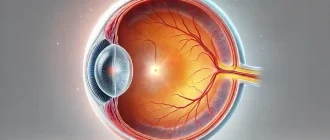
What is gonioscopy? Gonioscopy is carried out during the eye test to examine the internal drainage system of the eye, also referred to as the anterior chamber angle. The “angle” is where the cornea and the iris meet. This is the place where fluid inside the eye (liquid humor) drains out of the eye and into the venous system. Under normal circumstances, the angle can not be seen on test. A special contact lens prism placed on the surface area of the eye allows visualization of the angle and drainage system.
Why Is Gonioscopy Needed?
The pressure inside the eye is maintained by continuous production and drainage of fluid. If the drain system is not working appropriately, the pressure inside the eye, likewise referred to as intraocular pressure, can increase. High intraocular pressure can cause damage to the optic nerve, the “cable television” that sends images from the eye to the brain. This sort of damage is called glaucoma, the 2nd leading cause of blindness worldwide.
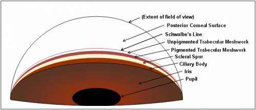
By taking a look at the “angle,” doctors can identify if it is open or closed in addition to if there are abnormal capillary, adhesions (synechiae), or damage from previous eye trauma. A closed angle is an abnormality that can predispose the patient to have a sudden or fast boost in intraocular pressure. This increase in pressure can cause an extremely severe, severe form of glaucoma that can be treated as well as prevented with laser treatment (iridotomy) if the predisposing angle abnormality is acknowledged using gonioscopy.
In addition, gonioscopy allows the eye doctor to note more subtle qualities of the eye’s drain system, in order to direct his or her medical diagnosis and treatment plan.
How Is Gonioscopy Done?
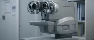
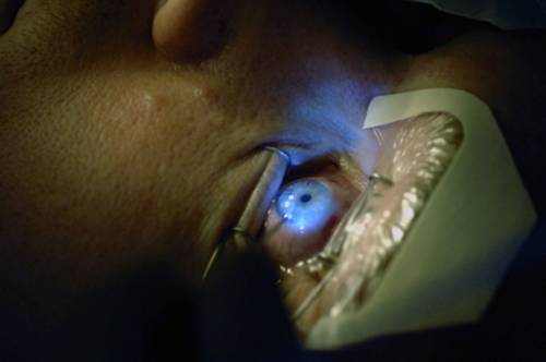
Gonioscopy is carried out with the head positioned in the slit lamp (the special microscope used to look at the eyes). After numbing the eye with drops, an unique contact lens is positioned straight on the eye and a beam is used to light up the angle. While the eyelids may feel the existence of the lens, there is normally no pain associated with this exam. Examination of both eyes typically takes a couple of minutes.
The Gonioscopy Process
Although the details vary based on the type of goniolens used, in basic the gonioscopy procedure includes:
- briefly explaining the procedure to the patient
- cleansing and sterilising the front (curved) surface area of the goniolens
- applying lubricating fluid to the front surface area if appropriate
- anaesthetising the patient’s cornea with topical anaesthetic
- preparing the slit light for viewing through the goniolens
- carefully moving the patient’s eyelids far from the cornea
- gradually applying the goniolens to the ocular surface, forming suction
- fine-tuning the slit light to optimise the view
- translating the gonioscopic image
- swivelling the goniolens to see each section of the iridocorneal angle
- when satisfied, really carefully breaking suction via the eyelids
- cleaning the instruments and watering the patient’s eyes with [saline] if preferred