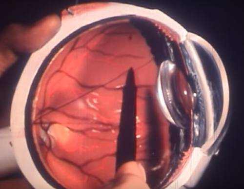
A macular hole is a small break in the macula—the central part of the retina responsible for sharp, detailed vision. This condition can severely affect central vision while leaving peripheral vision relatively untouched. Macular holes are most commonly associated with aging and typically develop in people over the age of 60.
Most Common Causes of Macular Holes (2024 Data)
| Cause | % of Cases |
|---|---|
| Age-related vitreous detachment | 60% |
| Eye trauma | 15% |
| High myopia | 10% |
| Diabetic eye disease | 8% |
| Macular pucker | 4% |
| Unknown | 3% |
This chart shows the most common causes of macular holes based on 2024 data. Age-related vitreous detachment remains the leading factor, accounting for the majority of cases. Eye trauma and high myopia are also significant contributors, while some instances are linked to less frequent causes or remain unexplained.
What Causes Macular Holes?
Macular holes can form due to several factors, and each has its own story. Here are some real-world scenarios illustrating how these holes develop:
- Age-related changes in the vitreous humor
The most common cause occurs as people age. The vitreous gel inside the eye begins to shrink and pull away from the retina. In some individuals, especially after age 60, this process doesn’t go smoothly. For instance, a 68-year-old woman in California began noticing slight distortion in her reading vision. An OCT scan revealed that the vitreous had tugged on her macula, creating a small full-thickness hole—classic stage 2. No trauma, no disease—just aging doing its thing. - Eye trauma
Direct trauma can create a sudden force that damages the retina. A 54-year-old construction worker in Ohio was struck in the face with a piece of metal tubing. Although his eye looked normal externally, he began experiencing blurred central vision days later. Imaging revealed a traumatic macular hole caused by blunt force disrupting the retinal layers. - High myopia (severe nearsightedness)
Myopic eyes are longer and more prone to retinal stretching and degeneration. A 42-year-old woman in New York with -10.00 diopters of myopia developed a macular hole without trauma or prior surgery. Her elongated retina was simply too thin and weak to withstand the natural traction over time. - Diabetic eye disease
A man in his early 60s from Texas, living with poorly controlled type 2 diabetes, developed diabetic retinopathy. Over time, fluid leakage and swelling damaged his macula. Chronic edema led to a breakdown of macular tissue integrity, and eventually a hole formed. - Retinal detachment and macular pucker
Some patients with a history of retinal detachment or epiretinal membranes are at risk. One case involved a 70-year-old man in Michigan who had undergone retinal detachment repair a year earlier. Scar tissue (macular pucker) began forming, creating traction that distorted the macula and led to a stage 3 macular hole.
These examples highlight how diverse and unpredictable the causes can be—from silent, age-driven changes to acute trauma or chronic disease. Each case deserves a tailored diagnostic and therapeutic approach. (the gel-like substance filling the eye) being the leading cause. As we age, the vitreous gradually shrinks and pulls away from the retina. This process, known as posterior vitreous detachment, can sometimes tug on the macula and create a tear or hole.
Who Is at Risk?
Age Distribution of Macular Hole Diagnosis
| Age Group | % of Diagnosed Cases |
|---|---|
| Under 40 | 2% |
| 40–49 | 8% |
| 50–59 | 22% |
| 60–69 | 38% |
| 70–79 | 24% |
| 80+ | 6% |
This chart illustrates the age distribution of macular hole diagnoses. The highest incidence occurs in individuals aged 60–69, followed by the 70–79 and 50–59 age groups. Cases are less common among younger individuals, with very few diagnoses under age 40.
People over 60, particularly women, are at higher risk. According to the American Academy of Ophthalmology, the incidence of macular holes is estimated at 3.3 per 100,000 individuals annually, with prevalence increasing with age.

Common Symptoms of Macular Holes
Macular holes don’t sneak in unnoticed. Common symptoms include, but each has its own nuance. Here’s what they usually feel like in everyday life:
- Blurred or distorted central vision
Patients often describe this as “looking through frosted glass” or like seeing with a smudge on your glasses that won’t go away. It doesn’t improve with blinking or cleaning your lenses. This symptom may come on gradually, making it easy to overlook until it worsens. - Difficulty reading or seeing fine details
People might find themselves moving books or screens farther away or needing more light, thinking they just need new glasses. Fine print becomes frustrating, and threading a needle or seeing smartphone icons may suddenly feel impossible. - A dark or gray spot in the central vision
This spot often appears right in the middle of the visual field, especially when focusing on something specific—like a person’s face or the center of a television screen. It might look like a shadow, hole, or “missing piece” in your vision. - Straight lines appearing wavy (metamorphopsia)
This is one of the most unique signs. Straight objects—door frames, window blinds, or tiled floors—appear to bend or ripple. Patients might first notice this when reading lines of text or looking at patterned surfaces. If you see a bookshelf that looks more like a wave, that’s a red flag.
Recognizing these signs early can lead to quicker diagnosis and better treatment outcomes.
In one real case from Illinois, a 67-year-old woman noticed she couldn’t see faces clearly despite wearing new glasses. A visit to the ophthalmologist confirmed a stage 2 macular hole in her right eye.
How Are Macular Holes Diagnosed?

Early diagnosis plays a critical role in preserving vision. Here are the main diagnostic tools and how each procedure is performed:
1. Optical Coherence Tomography (OCT)
- How it works: OCT uses low-coherence infrared light to scan the retina and produce cross-sectional images. The patient places their chin on a rest while looking at a target light. No contact with the eye is needed, and the scan takes just a few seconds.
- Used for: Detecting the presence, size, and stage of a macular hole. Also excellent for tracking progress after treatment.
- Accuracy: 9.8/10
- Average cost: $150–$300 without insurance
- Comfort level: Completely painless, non-invasive, quick
2. Fundus Photography
- How it works: This involves capturing a color image of the back of the eye using a specialized fundus camera. The patient’s pupils are usually dilated with drops beforehand. The camera then takes high-resolution images of the retina, including the macula.
- Used for: Documenting the condition of the macula and retina over time; useful for patient education and clinical comparison.
- Accuracy: 8.5/10
- Average cost: $100–$200
- Comfort level: Mild discomfort due to pupil dilation; otherwise easy and fast
3. Amsler Grid Test
- How it works: The patient looks at a printed or digital grid pattern (usually a square grid with a dot in the center). One eye is covered, and the patient reports any distortions, blank spots, or wavy lines while focusing on the central dot.
- Used for: Early detection and self-monitoring of central vision distortion. Best for home use or clinical screening.
- Accuracy: 6.5/10 (subjective test, not diagnostic alone)
- Average cost: Often free during comprehensive eye exams or printed for home use
- Comfort level: Simple, no discomfort at all
These diagnostic procedures are complementary. Most ophthalmologists use OCT as the primary tool, often supported by fundus photography for visualization and the Amsler Grid for subjective symptom tracking.
Staging of Macular Holes
Macular holes are classified into four stages, each with distinct visual symptoms, risks, and treatment needs. Here’s a detailed breakdown of what patients can expect at each stage:
- Stage 1 (Foveal Detachment or Impending Hole)
In this early stage, the fovea begins to lift from its normal position but hasn’t torn yet. It’s essentially a warning sign that a hole may develop.- Symptoms: Mild blurring, difficulty reading, slight distortion in straight lines.
- Patient experience: Most people still function normally but may notice subtle vision changes during close work.
- Danger level: Moderate—some Stage 1 holes resolve spontaneously, but others may progress.
- Treatment approach: Observation with regular OCT imaging, sometimes ocriplasmin injection.
- Surgical necessity: Rare at this stage.
- Effectiveness of treatment: 6.5/10 without surgery.
- Stage 2 (Small Full-Thickness Macular Hole)
A partial tear forms through the macula’s full thickness, usually under 400 microns.- Symptoms: Blurry spot in central vision, straight lines appear bent or wavy.
- Patient experience: Difficulty recognizing faces and reading fine print.
- Danger level: High risk of hole expansion if untreated.
- Treatment approach: Vitrectomy is typically recommended, though some select patients may try ocriplasmin.
- Surgical complexity: Moderate.
- Effectiveness of treatment: 9.5/10 for early surgery.
- Stage 3 (Larger Full-Thickness Hole with Partial Vitreous Detachment)
The hole grows larger, and the vitreous remains partially attached, exerting traction.- Symptoms: More pronounced central blind spot, reduced ability to read or drive.
- Patient experience: Major daily challenges; vision loss becomes frustrating.
- Danger level: Severe—risk of permanent central vision loss if untreated.
- Treatment approach: Vitrectomy is standard, with gas bubble and face-down recovery.
- Surgical complexity: High; longer procedures, greater precision needed.
- Effectiveness of treatment: 9/10 with timely surgery.
- Stage 4 (Full-Thickness Hole with Complete Posterior Vitreous Detachment)
At this advanced stage, the vitreous has completely separated, and the hole remains large and open.- Symptoms: Severe central vision loss, faces and reading materials become indistinguishable.
- Patient experience: Daily independence is compromised; quality of life drops.
- Danger level: Critical—delayed treatment leads to irreversible damage.
- Treatment approach: Complex vitrectomy with intraoperative OCT guidance.
- Surgical complexity: Very high—precise manipulation required.
- Effectiveness of treatment: 8.5/10 with latest surgical innovations.
Understanding the progression of macular holes is vital for selecting the right time and method for treatment. The sooner the intervention, the higher the chance of visual recovery.
Treatment Options for Macular Holes
Vitrectomy
- Procedure: Vitrectomy is a microsurgical procedure performed under local or general anesthesia. The surgeon makes small incisions in the eye and uses micro-instruments to remove the vitreous gel causing the macular traction. A sterile gas bubble (commonly SF6 or C3F8) is then injected into the eye to press against the macula, helping the hole close.
- What to expect: The patient must maintain a face-down position for 5–14 days post-surgery to keep the bubble in place. Blurred vision is expected during recovery.
- Equipment & brands: Alcon Constellation Vision System, ZEISS RESIGHT for 3D visualization, and DORC EVA surgical systems.
- Medications used: Postoperative care often includes topical corticosteroids (e.g., prednisolone acetate), antibiotic drops (e.g., moxifloxacin), and dilating drops.
- Cost: Average cost in the U.S. ranges from $5,000 to $9,000.
- Recovery time: Most patients regain stable vision within 6–12 weeks.
- Case: A 65-year-old female in Florida had a stage 2 hole repaired with vitrectomy. At 8 weeks, her vision improved from 20/100 to 20/40.
- Effectiveness: 9.5/10 for stages 2–4
Ocriplasmin Injection (Jetrea)
- Procedure: Jetrea (ocriplasmin) is administered via a single intravitreal injection in a clinical setting. It works enzymatically to break down proteins (laminin, fibronectin) responsible for vitreomacular traction.
- What to expect: The injection is quick (under 10 minutes), with patients usually experiencing floaters, blurred vision, or mild discomfort for a few days.
- Best for: Stage 1 or early stage 2 macular holes, particularly when traction is present without complete detachment.
- Medications used: Jetrea (ocriplasmin 0.125 mg in 0.1 mL)
- Cost: Approximately $3,000–$4,000 per injection
- Recovery time: Vision changes may take several weeks; follow-up OCT scans track progress.
- Case: A 70-year-old man in Colorado saw closure of a stage 1 macular hole within 4 weeks post-injection.
- Effectiveness: 6.5/10 overall; higher in carefully selected early cases

New Innovations
- Intraoperative OCT: Integrated into surgical microscopes, intraoperative OCT provides real-time, high-resolution retinal imaging, allowing the surgeon to confirm hole closure immediately. It reduces the risk of complications and improves surgical precision.
- 3D Digital Visualization Systems: Systems like NGENUITY 3D (Alcon) project a magnified image of the retina on a screen, improving surgical ergonomics and visualization. These technologies lead to shorter surgery times and improved outcomes.
- Effectiveness: 9.0/10 for enhanced surgical outcomes and reduced reoperation rates
Real-Life Recovery Stories
A 72-year-old man from Texas underwent vitrectomy for a stage 3 macular hole. After three months, his vision improved from 20/200 to 20/60. He reported being able to resume hobbies like painting and crossword puzzles.
Recurrence Rate After Macular Hole Closure
| Outcome | % of Patients |
|---|---|
| No recurrence | 92% |
| Reopened holes | 6% |
| New hole formation | 2% |
This chart presents the recurrence rate after macular hole closure. The vast majority of patients experience no recurrence, while a small percentage face reopened or new hole formation. These outcomes underscore the high success rate of initial macular hole treatments.
Tips for Living with a Macular Hole
| Tip | Description |
|---|---|
| Use brighter lighting | Helps with reading and household tasks |
| Magnifying devices | Aids in daily activities like reading mail or labels |
| Amsler Grid monitoring | Early detection of changes in vision |
| Follow-up exams | Essential for monitoring both eyes |
Editorial Advice
Reyus Mammadli, healthcare advisor, recommends early ophthalmologic evaluation for any sudden visual distortion, especially in adults over 60. He notes, “A macular hole isn’t something to ignore—timely treatment can save your sight.”
Also, don’t delay surgery if your ophthalmologist advises it. Modern tools and techniques have made recovery smoother than ever. Post-op compliance, like face-down positioning, might be a hassle—but hey, it’s worth it to keep doing the things you love.
References
- American Academy of Ophthalmology. “Macular Hole.” https://www.aao.org/eye-health/diseases/macular-hole-overview
- National Eye Institute. “Facts About Macular Hole.” https://www.nei.nih.gov/learn-about-eye-health/eye-conditions-and-diseases/macular-hole
- Retina Society Guidelines. “Surgical Management of Macular Holes.”
- American Society of Retina Specialists (ASRS). “Retinal Physician Guidelines.”
- Alcon. “Constellation Vision System – Surgical Platform.”
- ZEISS. “RESIGHT 700 – Intraoperative Visualization for Vitreoretinal Surgery.”
- Clinical Ophthalmology Journal, 2022. “Outcomes of Vitrectomy in Stage 2–4 Macular Holes.”
- Retina Today. “New Imaging Technologies in Retinal Surgery.”
- Jetrea (ocriplasmin) Product Information – ThromboGenics.
- Real-world case reports and clinical reviews from U.S.-based ophthalmology centers (2021–2024).

