Although it is little, the eye is a complicated organ. To enable clear vision, all structures within the eye should work effectively in order to capture light, focus it, and relay messages back to the brain to produce a visual image. This intricacy is what makes eye anatomy such a fascinating subject.
When we are born our eyes are only 1.6 to 1.7 centimeters in diameter. Over the first 3 years of life, the eyes grow rapidly, reaching their full size (simply shy of one inch, or 2.4 cm) by the age of 13. The visible part of the eyeball comprises 1/6 of the eye’s overall area, with the rest hidden behind the eyelids.
There are several physical and chemical elements that make up the eye. The eye is likewise greatly included with the nerve system, which enables the brain to take in info from the eyes and make the suitable choices on how to act on this information. The nerves need to be kept in prime condition or the brain might start to get false images, or you will not take in enough info to obtain a precise perception of your environment.
How the Eye Works
The eye is a complicated maker with numerous parts. It allows you not just to see items, however to see depth, color, size, and detail. The eye works by refracting and focusing light onto the retina. When light strikes the retina, countless rhodopsin-containing rods, which are responsible for night vision, transform the light into electrical impulses, which are sent out to the brain.
The brain then equates what it receives from the optic nerves so that we can comprehend what we see. The retina also consists of millions of cones that contain iodopsin and are used for bright light vision and color understanding. There are roughly 17 times more rods than cones– about 120 million rods and 7 million cones– in the retina of each eye.
Vision Support: Quality AREDS 2 Formula eye vitamins can be found on Amazon.
Parts of the Eye
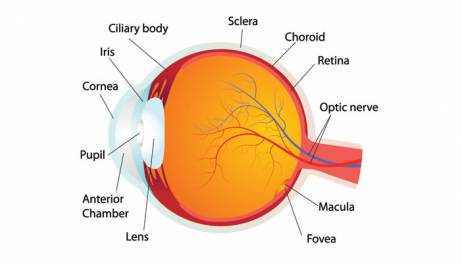
The detailed anatomy of the eye permits light refraction, preserves the shape of the eye, converts light into electrical impulses, and far more. Here is a look at the various parts of the eye:
The Cornea
The cornea is the dome-shaped outer covering of the eye. It resembles the windows of the car we discussed previously. It protects your eye and permits you to see around you. The cornea is where light is focused.
It comprises many layers, consisting of the external layer, the epithelium. The epithelium is typically gotten rid of or cut during surgical procedures that improve the cornea to focus light much better. Unlike other organs in the human body, there are no capillary in the cornea, given that capillary obstruct light from getting in the eye. Rather, the cornea gets its oxygen and nutrients from tears, from the atmosphere, and from the liquid humor.
Expert Choice: Recommended by eye care professionals for managing dry eyes, styes, and blepharitis, this Heated Eye Mask provides the consistent moist heat therapy endorsed by the American Academy of Ophthalmology. It’s an effective, reusable solution for soothing irritation and improving oil gland function. Available on Amazon.
- Main refractive surface area of the eye
- Index of refraction: n = 1.37
- Usually transparent and uniformly thick
- Almost avascular
- Richly supplied with nerve fibers
- Conscious foreign bodies, cold air, chemical inflammation
- Nutrition from aqueous humor and
- Tears maintain oxygen exchange and water content
- Tears prevent scattering and improve optical quality
The Sclera
The Sclera is the white outer part of the eye that you can see. It supplies protection and structure for the inner parts of the eye.
- Exterior is smooth and white
- Interior is brown and grooved
- Incredibly durable
- Versatility includes strength
- Constant with sheath of optic nerve
- Tendons attached to it
The Conjunctiva and Lacrimal Glands
The conjunctiva is a mucus layer that keeps the eye moist. It covers the sclera and the inner surfaces of the eyelids. Infections in this area are typically referred to as “Pink Eye.” Lacrimal glands, which produce tears, are discovered on the outer part of each eye.
The Vitreous Humor and Liquid Humor
Vitreous humor makes up roughly 80 percent of the volume of the eyeball. It is a gel-like compound in the back part of the eye that offers the shape of the eyeball. The vitreous humor lies between the lens and the retina, in an area called the vitreous cavity.
- Fills the space between lens and retina
- Transparent gelatinous body
- Particular viscosity of 1.8 – 2.0 (jelly-like consistency).
- Index of refraction, n= 1.33.
- Nutrition from retinal vessels, ciliary body, liquid.
- Floaters, shadows of sloughed off material/debris in the vitreous.
- Also keeps eye shape.
Besides helping to preserve the shape of the eyeball, the vitreous cavity also provides a clear pathway for light going through the eye to the retina. The Liquid Humor is the watery region in the front of the eyeball.
Specialized Care: For persistent itching or flaking, eye care specialists recommend targeted formulas like this BetterLids Eyelid Ointment . This preservative-free, dermatologist-tested oat complex is a top choice for soothing sensitive skin and managing symptoms of blepharitis. Available for easy ordering on Amazon.
It is separated into two regions, the anterior chamber in front of the iris, and the posterior chamber behind it. The canal of Schlemm drains pipes water in this region. Blockage of this canal causes glaucoma and other complications.
The main function of the liquid humor is to carry nutrients to the cornea and the lens and to get rid of waste products from inside the front of the eye via the canal of Schlemm.
The Iris and Pupil
The pupil is the great void in the center of the colored iris. It contracts when exposed to intense light and expands in darkness to permit more light into the eye. The iris is the colored part of the eye. This coloring is because of pigment cells in the tissue.
People with blue eyes have less pigment in their iris than those who have brown eyes. The iris includes the sphincter pupillae, the muscle used to narrow the pupil, and the dilator pupillae, the muscle used to widen it. The iris controls how much light gets in the eye by blocking extraneous light from getting in the student.
- Iris is greatly colored
- Sphincter muscle to restrict or dilate the pupil
- Pupil is the hole through which light passes
- Pupil diameter varies from about 3-7 mm
- Area of 7-38 square mm (aspect of 5).
- Eye color (brown, green, blue, etc.) dependent on amount and circulation of the pigment melanin.
The Lens
The lens is a clear structure behind the student that does just what a routine lens does. The primary purpose of the lens is to focus light by altering shape. The ciliary body is a muscle group attached to the lens that help the lens alter its shape to better focus light on the retina. As we age, our lenses naturally deteriorate, often resulting in cataracts.
- Transparent body enclosed in a flexible pill
- Made up of proteins and water
- Include layers, like an onion, with firm nucleus, soft cortex
- Gradient refractive index (1.38 – 1.40).
- Young person can change shape of the lens through ciliary muscles.
- Contraction of muscle triggers lens to bulge.
- At roughly age 50, the lens can no longer change shape.
- Becomes more yellow with age: Cataracts.
The Retina
The retina is the innermost layer of sensitive tissue that transfers light to the brain. The retina includes numerous types of cells, including a layer of rods and cones, which transform light into chemical and electrical energy that is sent to the optic nerves.
The center of the retina contains the macula. The macula is an extremely sensitive part of the retina that is responsible for our information vision. The center of the macula, which has a major role in information understanding, is called the fovea. When there is damage to the macula, we are unable to see fine details.
The Macula and Fovea
The macula is the center portion of the retina. Its main function is to offer clear, unique main vision. The fovea is the center part of the macula that provides the sharpest vision. The fovea just contains cones. Damage to the macula or fovea often results in a decrease in one’s central vision.
Covering the fovea is a pigment called the macula. it is thought that the macula serves as a protective filter over the fovea that absorbs blue and ultraviolet radiation. This pigment varies from observer to observer and is a source of specific variation in color vision. Normally we do not discover the filtering of the macula however under unique conditions we can discover its presence triggering what is known as Maxwell’s spot.
Here is a plot of the density of the macula as a function of wavelength:
To see Maxwell’s spot attempt at the same time viewing through a blue and yellow filter. When taking a look at through the blue filter after adjusting through the yellow filter you may see a dark region covering around the central 3° of visual angle.
The middle- and long- wavelength delicate cones are selectively adjusted to the yellow so that their response is attenuated while consequently looking through the blue, thus improving the visual impact of the macula.
Another demonstration of the macula is called Haidinger’s Brushes.
Take a look at a consistent blue field (again the clear sky works well for this) through a direct polarizer. You might be able to see a small yellow hourglass in the central 3° area. As you change the orientation of the polarizer, the orientation of the hour glass changes.
To the right is an artists depiction of Haidinger’s Brushes.
The fovea is the area on the retina of central look. When you look directly, or fixate, at a stimulus you the retinal locus of this central fixation is the fovea. There are only cones in the human fovea (no rods). They are thinner, lengthened, any very firmly loaded. Due to the fact that of this, the fovea is the area of greatest visual skill and best color vision.
In the diagram below you can see that the retinal layers are pulled aside (the axons of the receptors are extended) leaving a clearer course for the light to reach the receptors. There is really a little indentation or pit at the location of the fovea due to this and it is a clear landmark in the retina during an ophthalmic examination. The elongated external sectors of the cones (where the photopigment is and where the transduction occurs) increase the level of sensitivity by increasing the amount of photopigment. There is no vasculature in the central fovea.
The Optic Nerve
Also referred to as Cranial Nerve 2, the optic nerve is what carries messages from the eye to the brain. It consists of over one million axons, which bring visual details to various parts of the brain.
The Choroid
Located between the retinal pigment epithelium (see below) and the back wall of the eye, the choroid brings nutrients to the retina and the retinal pigment epithelium. The choroid is made up of melanin, which absorbs any extraneous light that might interfere with the image the eye is sending to the brain.
The Retinal Pigment Epithelium
The retinal pigment epithelium can be found between the retina and the choroid. The retinal pigment epithelium:
- Safeguards the retina from excess inbound light
- Products omega 3 fatty acids for building photoreceptive membranes
- Products glucose for energy
- Assists transport water from the retina to the choroid
- Keeps the pH balance of the retina
- Assists eliminate dead sections of photoreceptor cells
- Secretes compounds to help develop and sustain the choroid and retina.
Peripheral Eye Anatomy
There are other aspects of eye anatomy besides the eye itself, consisting of the eye socket or orbit, and the muscles that move the eye.
The Eye Muscles
The eyes have four groups of muscles:
- The extra-ocular muscles that manage eye motion. Each eye has six of these muscles, which manage each eye’s motion, permitting both eyes to see the same image at the same time.
- The muscles of the iris. These dilate and constrict the student of the eye, managing how much light enters it.
- The eyelid muscles that control opening and closing of the lids.
- The ciliary muscles. These control lens focusing within the eye.
The Orbit
The orbit is the pocket of tissue each eyeball sits in. Seven separate facial bones produce the walls around the orbit. Besides the eyeball, numerous muscles, nerves, blood vessels, fat, and the lacrimal drain system develop the complex structure.The optic nerve rests at the back of the orbit.
The Eyelids
The eyelids’ primary function is to protect the eyes by blinking. Blinking prevents debris from entering the eye. The typical blink rate is 10 blinks per minute. Males and female blink at about the exact same rate unless the woman is taking contraceptive pills; she’ll then blink at about 14 blinks per minute.
When an individual is focusing on reading or working on a computer, they’ll blink about 3 or four times a minute. This is the significant factor that eyes dry and end up being fatigued when reading.
The Lacrimal Drainage System
As mentioned above, the lacrimal glands, which are part of the drain system, likewise produce tears. The lacrimal drain system functions by distributing those tears over the surface area of the eye and getting rid of excess tears.
The puncta consist of little holes that allow tears to drain pipes from the eyes into the nose. If you were to divide your eyelids into thirds vertically, you would see that the innermost third of both the upper and lower lids includes the puncta.
The lacrimal drain system also contains the nasolacrimal sac and the nasolacrimal duct. The sac is a pouch situated under the skin in between the eye and the nose. Its primary function is to gather tears leaving the eye and ensure that they continue their course from the eye and into the nose. The duct is a tube that transfers the tears from the eye to the sac to the nose.
The Tear Film
Also part of the lacrimal drain system, tears are made from three elements: water, lipid, and mucus. Once they are produced from the lacrimal gland, they bathe the surface of the eye. Tears supply wetness and nutrition for the cornea and eliminate surface debris.
Once they have performed their tasks, they enter the puncta and travel through the nasolacrimal sac and duct, making their way into the nose and down the throat. As you can see, the eye is little however really intricate. So look after your eyes. Visit your eye care expert routinely, or if changes occur in your vision.
Did you understand … the eye can seeing a candle light flame from more than thirty miles away? Did you understand … messages are sent from the eye to the brain through the optic nerve at a speed of 423 miles per hour?