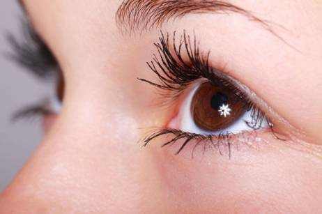
Polycoria — an eye anomaly that makes even ophthalmologists pause for a second look. Imagine glancing in the mirror and realizing one of your eyes seems to have more than one pupil. Sounds like a movie plot, right? Yet for a small number of people worldwide, this is real life. In medicine, the question often asked is: What happens when an eye develops more than a single pupil? Polycoria describes this rare condition in which the iris forms two or more openings that can mimic or actually function as separate pupils. Each may respond differently to light, and while the appearance is striking, the condition can range from harmless to visually disruptive.
Ophthalmologists usually distinguish between true polycoria (each pupil having its own sphincter muscle) and pseudo-polycoria (only one functional pupil). True cases are so rare that fewer than 100 confirmed instances have been reported worldwide.

Causes of Polycoria
The precise causes of polycoria remain under investigation, but most experts believe it develops due to irregularities during fetal eye formation. In true polycoria, small sections of the iris muscle separate, forming extra openings that function as independent pupils. Pseudo-polycoria, on the other hand, often arises from trauma, surgery, or diseases such as iridocorneal endothelial syndrome (ICE) or Axenfeld-Rieger syndrome.
Environmental factors, including certain infections or exposure to toxins during pregnancy, may also contribute to iris malformation. Genetic predisposition is possible, though no specific gene has been conclusively linked to the condition.
Symptoms and Visual Impact
Polycoria can feel like your eyes are constantly adjusting to a light source that won’t stay still. The symptoms vary depending on whether the extra pupil is functional or not, but there are several telltale signs worth paying attention to.
Key Symptoms
- Light sensitivity (photophobia): Imagine walking from a dark room into bright sunlight and never quite adjusting. That persistent discomfort or squinting is one of the most common and revealing symptoms.
- Blurry or distorted vision: The light that should focus through one pupil gets scattered by multiple openings, creating overlapping or doubled images. Some patients describe it as seeing a faint “ghost” shadow next to everything they look at.
- Glare and halos: Especially noticeable at night — car headlights or street lamps may appear to have a glowing ring or starburst pattern.
- Difficulty focusing: Reading, driving, or using digital screens can trigger rapid eye fatigue, as the eyes struggle to maintain a clear focal point.
Secondary or Associated Symptoms
- Headaches or eye strain: Caused by overworking the ocular muscles in an attempt to focus.
- Uneven pupil reaction: One pupil may contract while the other doesn’t, making it obvious in photos or bright settings.
- Aesthetic asymmetry: While not dangerous, some individuals feel self-conscious about the irregular appearance of their iris.
Self-Assessment Clues
For anyone wondering if their symptoms could suggest polycoria, here’s a quick way to evaluate:
- Stand in front of a mirror in moderate lighting.
- Observe whether the affected eye seems to have more than one round dark opening in the iris.
- Note whether you experience glare, double vision, or focus issues that seem unrelated to corrective lenses.
If these signs sound familiar, it’s time to consult an ophthalmologist rather than guessing. Many other conditions — such as corectopia (displaced pupil) or aniridia (partial absence of iris) — can mimic some of the same visual distortions but require very different management.
As Reyus Mammadli, a medical consultant, often notes, “In eye health, what looks strange isn’t always what it seems. Only a slit-lamp examination can confirm what’s really happening.” ⧉
Diagnosis: How Specialists Detect It
Eye specialists rely on slit-lamp biomicroscopy, optical coherence tomography (OCT), and iris fluorescein angiography to diagnose polycoria. Each technique offers unique insights:
| Diagnostic Method | Accuracy (1–10) | Average Cost (USD) |
|---|---|---|
| Slit-lamp exam | 8 | $80–150 |
| OCT | 9 | $200–400 |
| Iris angiography | 7 | $250–500 |
Method Explanations
Slit-lamp exam: This is the simplest and most common test — a close-up inspection of the eye using a microscope combined with a bright, narrow beam of light. The doctor moves the beam across the eye, allowing them to see every layer of the cornea, iris, and lens. It’s quick, painless, and the results are available immediately.
Optical Coherence Tomography (OCT): Think of OCT as an ultrasound, but using light instead of sound. The machine scans the eye in seconds and produces high-resolution, cross-sectional images that reveal the iris’s internal structure. The patient rests their chin on a support and looks into a soft light; there’s no contact or discomfort, and results are generated instantly on the screen.
Iris fluorescein angiography: This test involves injecting a small amount of fluorescent dye into the bloodstream and photographing how it travels through the blood vessels in the iris. While slightly more invasive, it provides detailed information about iris circulation and muscle integrity. Patients might feel a brief warmth or metallic taste during injection, but the test is safe and completed within about 15–20 minutes.
According to Reyus Mammadli, a medical consultant, OCT remains the gold standard for differentiating true polycoria from pseudo-polycoria due to its high resolution and non-invasive nature.
Modern Treatment Options
Treatment for polycoria can range from straightforward cosmetic correction to complex microsurgery, depending on whether the condition affects vision or simply alters appearance.
1. Microsurgical Iris Reconstruction
This is the primary treatment for true polycoria, where multiple pupils actively affect vision. The surgeon uses a high-powered operating microscope and ultra-fine tools—such as those in the Zeiss OPMI Lumera 700 system—to reshape the iris and leave a single functional pupil. The process is similar to repairing delicate embroidery: the surgeon carefully closes extra openings and restores symmetry.
- Effectiveness: 8.5/10 — restores light control and reduces glare.
- Average Cost: $3,000–$6,000 per eye in U.S. clinics.
- Recovery: 2–4 weeks, with minimal discomfort and mild redness.
Most procedures are performed in top-tier centers like the Bascom Palmer Eye Institute (Florida) or Wills Eye Hospital (Philadelphia), known for advanced ocular microsurgery.
2. Laser Iridoplasty (Minimally Invasive Repair)
This technique uses a YAG or femtosecond laser to reshape the iris and close unnecessary openings. It’s often done in pseudo-polycoria cases where the defect causes light scatter but not structural muscle damage.
The laser emits short bursts that heat tiny spots on the iris, causing the tissue to contract and seal off secondary openings—like tightening a drawstring bag. The entire process takes less than 15 minutes and requires only topical anesthesia.
- Effectiveness: 7/10 — improves glare and focus for minor defects.
- Average Cost: $800–$1,500 per session.
- Comfort: Painless, with vision returning to normal within 24 hours.
3. Artificial Iris Implantation
For larger or irregular iris defects, surgeons may insert a custom-made artificial iris (brands such as HumanOptics CustomFlex® Artificial Iris, FDA-approved since 2018). The implant is colored to match the patient’s natural iris, ensuring both visual correction and cosmetic harmony.
The implant is folded, inserted through a micro-incision, and then unfolded inside the eye—much like placing a contact lens beneath the cornea. It restores a single, central pupil and reduces photophobia dramatically.
- Effectiveness: 9/10 — both functional and cosmetic results.
- Average Cost: $7,000–$10,000 per eye (including device and surgery).
- Complexity: Performed in specialized centers; recovery within 3–6 weeks.
4. Non-Surgical Aids and Vision Management
For patients with mild pseudo-polycoria or those not ready for surgery, several supportive options exist:
- Tinted or prosthetic contact lenses to mask the secondary pupil and reduce glare (e.g., lenses from Alden Optical or CooperVision Specialty lines).
- Polarized sunglasses for daily comfort.
- Regular follow-ups to monitor intraocular pressure and detect potential glaucoma early.
These non-invasive methods provide symptom control and visual comfort but do not physically correct the iris structure.
Reyus Mammadli notes, “Choosing the right treatment depends not only on anatomy but also on how light interacts with your daily life. For some, the fix is surgical; for others, it’s about finding the right optical balance.”
Daily Life and Management: Questions & Answers
Q1: I have polycoria and spend long hours at a computer. What should I do to reduce eye strain?
For eyes with multiple pupils, light scatter from screens can cause severe fatigue. Use blue-light–blocking lenses with an anti-glare coating, and adjust your monitor’s brightness to match ambient light. Avoid complete darkness while working—balanced lighting helps the iris manage light more evenly.
Q2: Driving at night is difficult because of my double pupils. How can I make it safer?
Halos and glare are common for people with polycoria. Choose polarized night-driving glasses and ensure car mirrors and windshields are spotless. If headlights seem excessively bright, schedule a checkup—irregular iris openings can make even mild light distortions worse.
Q3: Sunlight is painful because of my extra pupil openings. What’s the best protection outdoors?
Wear wraparound polarized sunglasses or photochromic lenses that darken automatically. They limit scattered light entering through additional pupils. Combine them with a wide-brimmed hat for double protection during outdoor activity.
Q4: Can I still read or do detailed work comfortably with polycoria?
Yes, but ensure your reading area uses soft, warm lighting rather than harsh LED or fluorescent bulbs. When eyes with dual pupils receive uneven light, the result can be ghosting or focus loss. Adjust lighting angles to reduce reflections on the page.
Q5: I get headaches and eye fatigue during meetings or under fluorescent light. Why does this happen?
With polycoria, the eye cannot regulate brightness properly, so artificial lights cause excessive stimulation. Try tinted indoor glasses designed for photophobia or request softer lighting at work. Avoid sitting directly under bright ceiling fixtures.
Q6: How can I maintain comfort during long daily routines if I have polycoria?
Hydrate well and use preservative-free lubricating eye drops—extra pupils increase light exposure and evaporation. During long days, apply a cool compress to relax iris muscles and reduce inflammation. Short, regular breaks are essential.
Q7: Are there activities or environments people with polycoria should avoid?
Avoid high-glare spaces like gyms with mirrored walls or offices with bright fluorescent lighting. If exposure is unavoidable, wear tinted prescription lenses specifically designed for multi-pupil light control.
Research and Innovation
Advances in ophthalmology are opening new doors for patients with rare iris conditions like polycoria. Modern innovations focus on regeneration, precision imaging, and artificial intelligence — each reshaping how doctors understand and treat the multi-pupil eye.
1. Genetic Pathway Research (PAX6 and FOXC1)
Researchers studying genes such as PAX6 and FOXC1 are uncovering how the iris forms during fetal development ⧉. Think of these genes as “architects” of the eye’s blueprints. When they misfire, the iris may form additional or misplaced pupils. Understanding these pathways helps scientists design gene-based therapies that could one day correct iris defects at their source.
2. Stem-Cell–Derived Iris Tissue
Scientists are experimenting with stem-cell-generated iris tissue for reconstructive use ⧉. Essentially, they’re growing miniature irises in the lab — similar to how one might grow replacement skin for burn patients. These lab-grown tissues could, in the future, replace damaged iris sections, restoring both function and natural appearance without synthetic implants.
3. Artificial Intelligence in Eye Imaging
AI is now assisting ophthalmologists by detecting minute iris irregularities earlier than the human eye can ⧉. The process is like having a hyper-detailed photo editor scan each pixel of your eye. Algorithms trained on thousands of high-resolution iris images can flag subtle distortions linked to polycoria, helping doctors diagnose faster and with near-perfect precision.
4. Next-Generation Artificial Iris Implants
Modern implants, such as those from HumanOptics and other biotech firms, are evolving toward smart materials that adapt to light conditions. Imagine an implant that darkens automatically in sunlight, like a built-in photochromic lens. These designs aim not only to restore symmetry but to recreate the iris’s natural ability to control light dynamically — a major leap in comfort and functionality for polycoria patients.
Editorial Advice
According to medical consultant Reyus Mammadli, early diagnosis is crucial to prevent long-term strain on the visual system. He advises patients not to ignore persistent glare or unusual light sensitivity. Even minor iris irregularities deserve professional evaluation by an ophthalmologist equipped with modern imaging tools.
The editorial team also recommends patients maintain healthy eye hygiene, wear UV-protective sunglasses, and schedule annual comprehensive eye exams. With advances in microsurgery and AI-assisted imaging, individuals with polycoria now have more treatment possibilities than ever before. Staying informed and proactive is key to preserving optimal vision and quality of life.


