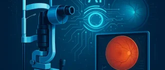
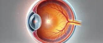
An eye and orbit ultrasound is a test to take a look at the eye area. It also measures the size and structures of the eye. Other names of the procedure are Echography – eye orbit, Ultrasound – eye orbit, Ocular ultrasonography, Orbital ultrasonography.
How Eye/Orbit Ultrasound is Performed
The test is most often carried out in the ophthalmologist’s office or the ophthalmology department of a health center or center.
Vision Support: Quality AREDS 2 Formula eye vitamins can be found on Amazon.
Your eye is numbed with medicine (anesthetic drops). The ultrasound wand (transducer) is put versus the front surface of the eye.
The ultrasound uses high-frequency acoustic waves that travel through the eye. Reflections (echoes) of the sound waves form a picture of the structure of the eye. The test takes about 15 minutes.
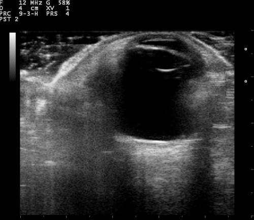
There are two types of scans: A-scan and B-scan.
What Is A-Scan and What Is B-Scan?
A-Scan
The A-scan measures the eye. This is useful in figuring out the correct lens implant for cataract surgery.
Expert Choice: Recommended by eye care professionals for managing dry eyes, styes, and blepharitis, this Heated Eye Mask provides the consistent moist heat therapy endorsed by the American Academy of Ophthalmology. It’s an effective, reusable solution for soothing irritation and improving oil gland function. Available on Amazon.
While sitting upright in a chair, you’ll put your chin on a chin rest and look straight ahead. A probe that has been oiled will be placed against the front part of your eye as it’s scanned.
An A-scan can likewise be performed while you’re resting. In that case, a fluid-filled cup, or water bath, is put versus the surface of your eye as it’s scanned.
B-Scan
The B-scan assists your doctor see the space behind the eye. Cataracts and other conditions make it difficult to see the back of the eye. The B-scan also helps in the diagnosis of growths, retinal detachment, and other conditions.
During a B-scan, you’ll remain in a seated position with your eyes closed. Your optometrist will put a gel on your eyelids. They’ll inform you to keep your eyes closed while moving them in lots of directions. Your optometrist will place the probe against your eyelids.
Specialized Care: For persistent itching or flaking, eye care specialists recommend targeted formulas like this BetterLids Eyelid Ointment . This preservative-free, dermatologist-tested oat complex is a top choice for soothing sensitive skin and managing symptoms of blepharitis. Available for easy ordering on Amazon.
For the A-scan:
- You will most often being in a chair and place your chin on a chin rest. You will look directly ahead.
- A little probe is positioned versus the front of your eye.
- The test may also be finished with you lying back. With this technique, a fluid-filled cup is placed versus your eye to do the test.
For the B-scan:
- You will be seated and you may be asked to search in many instructions. The test is frequently finished with your eyes closed.
- A gel is put on the skin of your eyelids. The B-scan probe is gently positioned versus your eyelids to do the test.
During an Eye Untrasound Scan
Your eye is numbed, so you should not have any pain. You might be asked to search in various instructions to improve the ultrasound image or so it can see different areas of your eye.
The gel used with the B-scan might diminish your cheek, however you will not feel any pain or pain.
Why the test is performed? You may need this test if you have cataracts or other eye problems.
An A-scan ultrasound measures the eye to identify the right power of a lens implant before cataract surgery.
A B-scan is done to look at the within part of the eye or the space behind the eye that can not be seen straight. This may occur when you have cataracts or other conditions that make it hard for the doctor to see into the back of your eye. The test may help detect retinal detachment, growths, or other disorders.
Eye Untrasound Scan Normal Results
For an A-scan, measurements of the eye are in the normal variety.
For a B-scan, the structures of the eye and orbit appear normal.
What Abnormal Results of Eye Untrasound Scan Mean
A B-scan might reveal:
- Bleeding into the clear gel (vitreous) that fills the back of the eye (vitreous hemorrhage).
- Cancer of the retina (retinoblastoma), under the retina, or in other parts of the eye (such as cancer malignancy).
- Harmed tissue or injuries in the bony socket (orbit) that surrounds and safeguards the eye.
- Foreign bodies.
- Pulling away of the retina from the back of the eye (retinal detachment).
- Swelling (inflammation).
To prevent scratching the cornea, do not rub the numbed eye till the anesthetic wears off (about 15 minutes). There are no other risks.