The retina is the transparent, light-sensitive structure at the back of the eye. The cornea and lens focus light onto the retina. The central area of the retina, called the macula, consists of a high density of color-sensitive photoreceptor (light-sensing) cells. These cells, called cones, produce the sharpest visual images and are responsible for central and color vision. The peripheral area of the retina, which surrounds the macula, consists of photoreceptor cells called rods, which respond to reduce light levels however are not color delicate. The rods are responsible for peripheral vision and night vision.
What Is Function of Healthy Retina?
The retina is a layer of tissue in the back of your eye that senses light and sends out images to your brain. In the center of this nerve tissue is the macula. It offers the sharp, main vision needed for reading, driving and seeing fine detail.
Vision Support: Quality AREDS 2 Formula eye vitamins can be found on Amazon.
Retinal disorders affect this vital tissue. They can affect your vision, and some can be severe sufficient to cause blindness.
A healthy retina is necessary for good vision. The eye is made up of light-sensitive cells connected with nerve fibers that allow light getting in the eye to be transformed to nerve impulses that reach the brain. The amount of light entering is controlled by the iris and is then passed to the retina. The retina is a thin membranous lining at the back of the eye.
It is light-sensitive nerve tissue that manages how images are seen. The images are focused here and converted to electrical impulses which are reached the brain by the optic nerve.
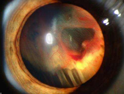
To take a look at the retina, an eye doctor will dilate, or expand, your eyes during a thorough assessment using eye drops. A retina expert then uses a special magnifying lens to examination your retina. The dilation will reverse after numerous hours.
Expert Choice: Recommended by eye care professionals for managing dry eyes, styes, and blepharitis, this Heated Eye Mask provides the consistent moist heat therapy endorsed by the American Academy of Ophthalmology. It’s an effective, reusable solution for soothing irritation and improving oil gland function. Available on Amazon.
List of Most Common Retina Problems
- Macular degeneration – a disease that damages your sharp, main vision
- Diabetic eye disease
- Retinal detachment – a medical emergency situation, when the retina is pulled away from the back of the eye
- Retinoblastoma – cancer of the retina. It is most common in children.
- Macular pucker – scar tissue on the macula
- Macular hole – a little break in the macula that typically takes place to people over 60
- Floaters – cobwebs or specks in your field of vision
The optic nerve carries signals created by the photoreceptors (cones and rods). Each photoreceptor is joined to the optic nerve by a small nerve branch. The optic nerve is connected to afferent neuron that bring signals to the vision center of the brain, where they are interpreted as visual images.
The optic nerve and the retina have a rich supply of blood vessels that bring blood and oxygen. Part of this supply of blood vessels originates from the choroid, which is the layer of capillary that lies in between the retina and the external white layer of the eye called the sclera. The central retinal artery (the other significant source of blood to the retina) reaches the retina near the optic nerve and then branches out within the retina. Blood drains from the retina into branches of the main retinal vein. The main retinal vein exits the eye within the optic nerve.
How to Detect Problems with Retina
When examining an individual’s retina, a doctor puts drops in the eye to dilate the student. This allows the retina to be seen in far more detail with ophthalmoscopy (shining a light through a magnifying lens and into the back of the eye).
Retinal disorders are typically identified and dealt with by an ophthalmologist. An ophthalmologist is a medical doctor who specializes in the evaluation and treatment (surgical and nonsurgical) of all types of eye conditions. Often, treatment is by an ophthalmologist who focuses on illness of the retina.
Specialized Care: For persistent itching or flaking, eye care specialists recommend targeted formulas like this BetterLids Eyelid Ointment . This preservative-free, dermatologist-tested oat complex is a top choice for soothing sensitive skin and managing symptoms of blepharitis. Available for easy ordering on Amazon.