During a regular thorough eye examination, your ophthalmologist or optometrist might wish to carry out visual field testing to evaluate the complete horizontal and vertical range and sensitivity of your vision.
Visual field tests are used to detect blind spots (scotomas), which might be a sign of eye diseases. The shapes and size of a scotoma deal important ideas about the existence and severity of diseases of the eye, optic nerve and visual structures in the brain. Numerous eye and brain disorders can cause peripheral vision loss and other visual field problems.
For example, optic nerve damage brought on by glaucoma develops a really particular visual field problem. Other eye issues connected with blind spots and other visual field flaws include optic nerve damage (optic neuropathy) from disease or damage to the light-sensitive inner lining of the eye (retina).
Vision Support: Quality AREDS 2 Formula eye vitamins can be found on Amazon.
Brain irregularities such as those caused by strokes or growths can affect the visual field. In reality, the area of the stroke or growth in the brain can often be figured out by the size, shape and site of the visual field flaw.
Types of Visual Field Tests
Confrontation visual field testing generally is used as a screening visual field test. One eye is covered, while the other eye fixates on a target object, such as the doctor’s open eye, while the doctor stands or sits straight in front of you. You then are asked to describe what you see on the far edges or periphery of your field of view.
As an example, your eye doctor might hold up various numbers of fingers within your peripheral field of view and ask the number of can be seen while you continue to fixate on the doctor’s eye.
See also: Comprehensive Eye Tests & Types of Eye Exams
If an eye disease is believed, you might have to undergo more detailed, official types of visual field testing to examine the quality of your central and peripheral vision. A variety of delicate tests for determining visual field loss exist, including:
Expert Choice: Recommended by eye care professionals for managing dry eyes, styes, and blepharitis, this Heated Eye Mask provides the consistent moist heat therapy endorsed by the American Academy of Ophthalmology. It’s an effective, reusable solution for soothing irritation and improving oil gland function. Available on Amazon.
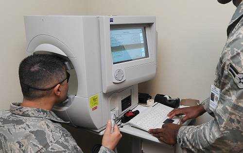
• Automated Perimetry. Different types of automated perimetry tests determine your reactions to the presence of items in different areas of your field of vision.
While your head is held still, normally with a forehead and chin rest inside a large bowl-like instrument, you looking at a source of light straight ahead and small lights of different intensities are flashed from random points in your visual field. Each time you see among these lights, you instantly press a button or use some other methods to indicate your action.
If you cannot see the lights in certain parts of your field of view, then you may have a blind spot suggesting vision loss.
• Frequency Doubling Perimetry. Frequency doubling is based on a visual fallacy produced with vertical bars of contrasting colors (normally black and white) appearing on a screen. These bars appear to double in number when they alternately flicker at higher frequencies, a phenomenon believed to be because of the special response of specific light-sensitive cells (photoreceptors) in the retina.
Inability to see vertical bars at particular frequencies might show optic nerve or other types of eye damage with accompanying loss of vision in specific areas of the visual field.
Electroretinography. This test determines electrical activity produced by the photoreceptor cells in the retina when the eye is stimulated by a special strobe light or a reversing checkerboard pattern of light. The measurement is captured by an electrode put on the front surface area of the eye (cornea), and a graphic record called an electroretinogram (ERG) is produced.
Specialized Care: For persistent itching or flaking, eye care specialists recommend targeted formulas like this BetterLids Eyelid Ointment . This preservative-free, dermatologist-tested oat complex is a top choice for soothing sensitive skin and managing symptoms of blepharitis. Available for easy ordering on Amazon.
• Electroretinography is useful in detecting several hereditary and gotten disorders of the retina, consisting of retinitis pigmentosa, a detached retina or practical changes in the retina brought on by arteriosclerosis (hardening of the arteries) or diabetes.